-
抗体試薬
- フローサイトメトリー用試薬
-
ウェスタンブロッティング抗体試薬
- イムノアッセイ試薬
-
シングルセル試薬
- BD® AbSeq Assay
- BD Rhapsody™ Accessory Kits
- BD® OMICS-One Immune Profiler Protein Panel
- BD® Single-Cell Multiplexing Kit
- BD Rhapsody™ TCR/BCR Next Multiomic Assays
- BD Rhapsody™ Targeted mRNA Kits
- BD Rhapsody™ Whole Transcriptome Analysis (WTA) Amplification Kit
- BD® OMICS-Guard Sample Preservation Buffer
- BD Rhapsody™ ATAC-Seq Assays
- BD® OMICS-One Protein Panels
-
細胞機能評価のための試薬
-
顕微鏡・イメージング用試薬
-
細胞調製・分離試薬
-
- BD® AbSeq Assay
- BD Rhapsody™ Accessory Kits
- BD® OMICS-One Immune Profiler Protein Panel
- BD® Single-Cell Multiplexing Kit
- BD Rhapsody™ TCR/BCR Next Multiomic Assays
- BD Rhapsody™ Targeted mRNA Kits
- BD Rhapsody™ Whole Transcriptome Analysis (WTA) Amplification Kit
- BD® OMICS-Guard Sample Preservation Buffer
- BD Rhapsody™ ATAC-Seq Assays
- BD® OMICS-One Protein Panels
- Japan (Japanese)
-
Change country/language
Old Browser
Looks like you're visiting us from United States.
Would you like to stay on the current country site or be switched to your country?
BD Pharmingen™ PE Mouse anti-Human IL-17F
クローン O33-782 (RUO)

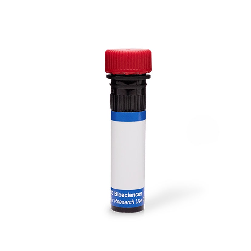
Flow cytometric analysis of IL-17F expression by resting and activated human peripheral blood CD4+ T cells and Th17 polarized cells. Human peripheral blood mononuclear cells were either unstimulated (Left Panels) or stimulated with Phorbol 12-Myristate 13-Acetate (PMA; Sigma P-8139) plus Ionomycin (Sigma; I-0634) in the presence of BD GolgiStop™ Protein Transport Inhibitor (Cat. No. 554724) for 5 hours (Middle Panels) or were cultured in Th17 polarization conditions and restimulated with PMA and Ionomycin in the presence of BD GolgiStop™ for 5 hours (Right Panels). Cells were then fixed and permeabilized using BD Cytofix/Cytoperm™ reagents (Cat. No. 554714) followed by staining with PE Mouse anti-Human IL-17F (Cat. No. 561197/561198), PerCP-Cy5.5 Mouse anti-Human CD4 (Cat. No. 341654), and FITC Mouse Anti-Human IFN-γ (Cat. No. 554700) or Alexa Fluor® 647 Mouse anti-Human IL-17A (Cat. No. 560490). Two-color flow cytometric dot plots showing the correlated expression patterns of IL-17F versus IL-17A or IFN-γ were derived from CD4+ gated events with the forward and side light-scatter characteristics of intact lymphocytes. Flow cytometry was performed using a BD™ LSR II Flow Cytometer System. Other compatible fixation and permeabilization treatments are listed in the \"Recommended Assay Procedure.\"

Flow cytometric analysis of IL-17F expression by resting and activated human peripheral blood CD4+ T cells and Th17 polarized cells. Human peripheral blood mononuclear cells were either unstimulated (Left Panels) or stimulated with Phorbol 12-Myristate 13-Acetate (PMA; Sigma P-8139) plus Ionomycin (Sigma; I-0634) in the presence of BD GolgiStop™ Protein Transport Inhibitor (Cat. No. 554724) for 5 hours (Middle Panels) or were cultured in Th17 polarization conditions and restimulated with PMA and Ionomycin in the presence of BD GolgiStop™ for 5 hours (Right Panels). Cells were then fixed and permeabilized using BD Cytofix/Cytoperm™ reagents (Cat. No. 554714) followed by staining with PE Mouse anti-Human IL-17F (Cat. No. 561197/561198), PerCP-Cy5.5 Mouse anti-Human CD4 (Cat. No. 341654), and FITC Mouse Anti-Human IFN-γ (Cat. No. 554700) or Alexa Fluor® 647 Mouse anti-Human IL-17A (Cat. No. 560490). Two-color flow cytometric dot plots showing the correlated expression patterns of IL-17F versus IL-17A or IFN-γ were derived from CD4+ gated events with the forward and side light-scatter characteristics of intact lymphocytes. Flow cytometry was performed using a BD™ LSR II Flow Cytometer System. Other compatible fixation and permeabilization treatments are listed in the \"Recommended Assay Procedure.\"

Flow cytometric analysis of IL-17F expression by resting and activated human peripheral blood CD4+ T cells and Th17 polarized cells. Human peripheral blood mononuclear cells were either unstimulated (Left Panels) or stimulated with Phorbol 12-Myristate 13-Acetate (PMA; Sigma P-8139) plus Ionomycin (Sigma; I-0634) in the presence of BD GolgiStop™ Protein Transport Inhibitor (Cat. No. 554724) for 5 hours (Middle Panels) or were cultured in Th17 polarization conditions and restimulated with PMA and Ionomycin in the presence of BD GolgiStop™ for 5 hours (Right Panels). Cells were then fixed and permeabilized using BD Cytofix/Cytoperm™ reagents (Cat. No. 554714) followed by staining with PE Mouse anti-Human IL-17F (Cat. No. 561197/561198), PerCP-Cy5.5 Mouse anti-Human CD4 (Cat. No. 341654), and FITC Mouse Anti-Human IFN-γ (Cat. No. 554700) or Alexa Fluor® 647 Mouse anti-Human IL-17A (Cat. No. 560490). Two-color flow cytometric dot plots showing the correlated expression patterns of IL-17F versus IL-17A or IFN-γ were derived from CD4+ gated events with the forward and side light-scatter characteristics of intact lymphocytes. Flow cytometry was performed using a BD™ LSR II Flow Cytometer System. Other compatible fixation and permeabilization treatments are listed in the \"Recommended Assay Procedure.\"


BD Pharmingen™ PE Mouse anti-Human IL-17F

Regulatory Statusの凡例
Any use of products other than the permitted use without the express written authorization of Becton, Dickinson and Company is strictly prohibited.
Preparation and Storage
推奨アッセイ手順
This antibody conjugate is suitable for intracellular staining of human peripheral blood mononuclear cells using BD Cytofix/Cytoperm™ reagents, BD Pharmingen™ Human FoxP3 Buffer Set or BD™ Phosflow fixation and permeabilization buffers (Fix buffer I with Perm/Wash Buffer I, Perm Buffer II, or Perm Buffer III).
Product Notices
- This reagent has been pre-diluted for use at the recommended Volume per Test. We typically use 1 × 10^6 cells in a 100-µl experimental sample (a test).
- An isotype control should be used at the same concentration as the antibody of interest.
- Source of all serum proteins is from USDA inspected abattoirs located in the United States.
- Caution: Sodium azide yields highly toxic hydrazoic acid under acidic conditions. Dilute azide compounds in running water before discarding to avoid accumulation of potentially explosive deposits in plumbing.
- For fluorochrome spectra and suitable instrument settings, please refer to our Multicolor Flow Cytometry web page at www.bdbiosciences.com/colors.
- Alexa Fluor® is a registered trademark of Molecular Probes, Inc., Eugene, OR.
- Cy is a trademark of GE Healthcare.
- Please refer to www.bdbiosciences.com/us/s/resources for technical protocols.
関連製品

最近閲覧済み
The O33-782 monoclonal antibody specifically binds to Interleukin-17F (IL-17F). IL-17F is a member of the IL-17 family of cytokines. IL-17F is encoded by the IL17F gene located in chromosome 6 (location: 6p12). IL-17F is a proinflammatory cytokine that is produced by activated T cells including differentiated CD4+ T helper 17 (Th17) cells. Activated Th17 cells can express disulfide-linked IL-17F and IL-17A homodimers as well as IL-17A/IL-17F heterodimers. These IL-17 dimers act by binding to and signaling through IL-17 receptor complexes (IL-17R). IL-17R are comprised of transmembrane IL-17RA and IL-17-RC protein subunits that are expressed by a variety of target cells including epithelial and endothelial cells, keratinocytes, fibroblasts, and granulocytes. IL-17F can induce target cells to produce proinflammatory cytokines such as IL-1β, IL-6, G-CSF, GM-CSF, and TNF and chemokines including CXCL1/Gro-α, CXCL2/Gro-β, and CXCL8/IL-8 that attract and activate leukocytes, eg, neutrophils. Th17 and other IL-17F-producing cells play protective roles in the clearance of extracellular pathogens, including bacteria and fungi. IL-17F can also play adverse roles in inflammation associated with asthma and autoimmune diseases.

Development References (6)
-
Fouser LA, Wright JF, Dunussi-Joannopoulos K, Collins M. Th17 cytokines and their emerging roles in inflammation and autoimmunity. Immunol Rev. 2008; 226:87-102. (Biology). View Reference
-
Shen F, Gaffen SL. Structure-function relationships in the IL-17 receptor: implications for signal transduction and therapy. Cytokine. 2008; 41(2):92-104. (Biology). View Reference
-
Starnes T, Robertson MJ, Sledge G, et al.. Cutting edge: IL-17F, a novel cytokine selectively expressed in activated T cells and monocytes, regulates angiogenesis and endothelial cell cytokine production. J Immunol. 2001; 167(8):4137-4140. (Biology). View Reference
-
Wang YH, Liu YJ. The IL-17 cytokine family and their role in allergic inflammation. Curr Opin Immunol. 2008; 20(6):697-702. (Biology). View Reference
-
Wright JF, Bennett F, Li B, et al. The human IL-17F/IL-17A heterodimeric cytokine signals through the IL-17RA/IL-17RC receptor complex. J Immunol. 2008; 181(4):2799-2805. (Biology). View Reference
-
Wright JF, Guo Y, Quazi A, et al. Identification of an interleukin 17F/17A heterodimer in activated human CD4+ T cells. J Biol Chem. 2007; 282(18):13447-13455. (Biology). View Reference
Please refer to Support Documents for Quality Certificates
Global - Refer to manufacturer's instructions for use and related User Manuals and Technical data sheets before using this products as described
Comparisons, where applicable, are made against older BD Technology, manual methods or are general performance claims. Comparisons are not made against non-BD technologies, unless otherwise noted.
For Research Use Only. Not for use in diagnostic or therapeutic procedures.
