-
抗体試薬
- フローサイトメトリー用試薬
-
ウェスタンブロッティング抗体試薬
- イムノアッセイ試薬
-
シングルセル試薬
- BD® AbSeq Assay | シングルセル試薬
- BD Rhapsody™ Accessory Kits | シングルセル試薬
- BD® Single-Cell Multiplexing Kit | シングルセル試薬
- BD Rhapsody™ Targeted mRNA Kits | シングルセル試薬
- BD Rhapsody™ Whole Transcriptome Analysis (WTA) Amplification Kit | シングルセル試薬
- BD® OMICS-Guard Sample Preservation Buffer
- BD Rhapsody™ ATAC-Seq Assays
- BD Rhapsody™ TCR/BCR Next Multiomic Assays
-
細胞機能評価のための試薬
-
顕微鏡・イメージング用試薬
-
細胞調製・分離試薬
-
- BD® AbSeq Assay | シングルセル試薬
- BD Rhapsody™ Accessory Kits | シングルセル試薬
- BD® Single-Cell Multiplexing Kit | シングルセル試薬
- BD Rhapsody™ Targeted mRNA Kits | シングルセル試薬
- BD Rhapsody™ Whole Transcriptome Analysis (WTA) Amplification Kit | シングルセル試薬
- BD® OMICS-Guard Sample Preservation Buffer
- BD Rhapsody™ ATAC-Seq Assays
- BD Rhapsody™ TCR/BCR Next Multiomic Assays
- Japan (Japanese)
-
Change country/language
Old Browser
Looks like you're visiting us from {countryName}.
Would you like to stay on the current country site or be switched to your country?
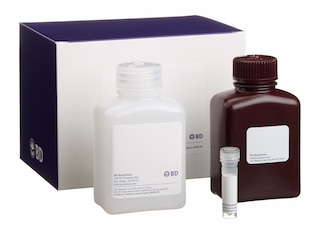
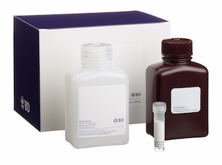
BD GolgiStop™ Protein Transport Inhibitor (Containing Monensin)
(RUO)

Regulatory Statusの凡例
Becton, Dickinson and Companyの書面による明示的な許諾を得た使用以外での製品の使用は固く禁じられています。
説明
The ex vivo addition of BD GolgiStop™, a protein transport inhibitor containing monensin, to in vitro- or in vivo-stimulated lymphoid cells blocks their intracellular protein transport processes. This results in the accumulation of cytokines and/or proteins in the Golgi complex. The increased accumulation of cytokines in the cell enhances the detectability of cytokine-producing cells with flow cytometric analysis.
Investigators should note that the appearance of BD GolgiStop™ may range in color from clear (colorless) to a light yellow.
調製と保管タイトルテキスト
推奨アッセイ手順
Stimulation of Cells: Various in vitro methods have been reported for stimulating cells to produce cytokines. Polyclonal activators have been particularly useful for inducing cytokine-producing cells. These activators include the following: concanavalin A, lipopolysaccharide, phorbol esters plus calcium ionophore or ionomycin, phytohaemaglutinin, staphlylococcus, entertoxin B, and monoclonal antibodies directed against subunits of the TCR/CD3 complex (with or without antibodies directed against costimulatory receptors, such as CD28).
Procedure for Using BD GolgiStop™: Add 4 µl of BD GolgiStop™ for every 6 mL of cell culture (e.g., ~10^6 cells/mL) and mix thoroughly. Treatment of stimulated cells for 4 to 6 hours with BD GolgiStop™ significantly increases the ability to detect cytokine-producing cells by immunofluorescent staining. It is recommended that BD GolgiStop™ not be kept in cell culture for longer than 12 hours.
As an alternative to BD GolgiStop™, investigators may wish to use BD GolgiPlug™ , a protein transport inhibitor containing brefeldin A (Cat. No. 555029). BD GolgiStop™ and BD GolgiPlug™ have been found to have differential effects on intracellular cytokine staining that is time, activator and cytokine dependent. These factors must be considered when carrying out intracellular staining.
Danger: BD GolgiStop™ Protein Transport Inhibitor, containing monensin (component 51-2092KZ) contains 99.61% ethanol (w/w) and 0.26% monensin, mononatriumsalz (w/w).
Hazard statements:
Highly flammable liquid and vapor.
Causes serious eye irritation.
Harmful if swallowed.
Precautionary statements:
Keep away from heat/sparks/open flames/hot surfaces. No smoking.
Wear protective gloves / eye protection.
Wear protective clothing.
IF ON SKIN (or hair): Remove / Take off immediately all contaminated clothing. Rinse skin with water / shower.
IF IN EYES: Rinse cautiously with water for several minutes. Remove contact lenses, if present and easy to do. Continue rinsing.
IF SWALLOWED: Call a POISON CENTRE/doctor if you feel unwell. Rinse mouth.
Dispose of contents / container in accordance with local / regional / national / international regulations.Keep container tightly closed.
製品通知
- Please refer to www.bdbiosciences.com/us/s/resources for technical protocols.
コンパニオン製品


Development References (5)
-
Assenmacher M, Schmitz J, Radbruch A. Flow cytometric determination of cytokines in activated murine T helper lymphocytes: expression of interleukin-10 in interferon-gamma and in interleukin-4-expressing cells. Eur J Immunol. 1994; 24(5):1097-1101. (Biology). View Reference
-
Elson LH, Nutman TB, Metcalfe DD, Prussin C. Flow cytometric analysis for cytokine production identifies T helper 1, T helper 2, and T helper 0 cells within the human CD4+CD27- lymphocyte subpopulation. J Immunol. 1995; 154(9):4294-4301. (Biology). View Reference
-
Jung T, Schauer U, Heusser C, Neumann C, Rieger C. Detection of intracellular cytokines by flow cytometry. J Immunol Methods. 1993; 159(1-2):197-207. (Methodology: Flow cytometry). View Reference
-
Prussin C, Metcalfe DD. Detection of intracytoplasmic cytokine using flow cytometry and directly conjugated anti-cytokine antibodies. J Immunol Methods. 1995; 188(1):117-128. (Methodology: Flow cytometry). View Reference
-
Sander B, Hoiden I, Andersson U, Moller E, Abrams JS. Similar frequencies and kinetics of cytokine producing cells in murine peripheral blood and spleen. Cytokine detection by immunoassay and intracellular immunostaining. J Immunol Methods. 1993; 166(2):201-214. (Biology). View Reference
Please refer to Support Documents for Quality Certificates
Global - Refer to manufacturer's instructions for use and related User Manuals and Technical data sheets before using this products as described
Comparisons, where applicable, are made against older BD Technology, manual methods or are general performance claims. Comparisons are not made against non-BD technologies, unless otherwise noted.
For Research Use Only. Not for use in diagnostic or therapeutic procedures.