細胞増殖には、サイトカイン処理など、数多くの刺激が影響を及ぼします。細胞増殖は、サイトカインへの曝露などの多くの刺激や、その他のさまざまなプロセスに反応して起こります。
Cell proliferation dyes
BD Biosciencesは、紫色レーザーで細胞増殖を検出するBD Horizon™紫色増殖色素450(VPD450)と、青色レーザーで細胞増殖を検出するBD Horizon™ CFSEを取り揃えています。これらの製品ではより大きなパネルが使いやすくなっています。そのため、マルチカラーフローサイトメトリーを用いて、限られたサンプルからより多くのデータを取得することができます。
いずれの増殖色素も非蛍光エステル型色素です。エステル基によって色素は細胞内に侵入することができます。色素が細胞内に侵入すると、エステラーゼはエステル基を切断して、色素を蛍光産物に変換し、細胞内でそれを捕捉します。複製イベントが発生する度に、細胞内の色素量が減少し、特徴的なパターンに至ります。

BD Horizon™細胞増殖色素は自由に細胞内に侵入することができます。細胞内に侵入すると、色素は非特異的エステラーゼによって切断され、蛍光分子を放出します。この蛍光分子が細胞内で捕捉されます。


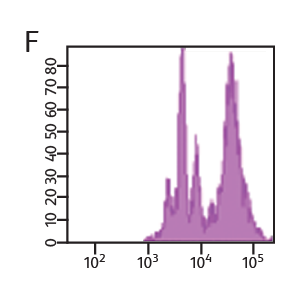
Concentration of VPD450 and cell-cycle kinetics on mouse spleen stimulated with anti-CD3e and anti-CD28. C57 Black/6 splenocytes were either loaded with varying concentrations of BD Horizon™ VPD450, DMSO, or left as untreated controls, then stimulated with anti-CD3e and anti-CD28 for two days. Cells were pulsed with BrdU prior to harvesting, then stained with APC anti-BrdU and 7-AAD (Cat. No. 552598). The top panels (A–C) illustrate APC antiBrdU and 7-AAD staining. The bottom panels (D–F) illustrate the corresponding VPD450 histograms. The control cells (Cells– ) (Panel A) and the 1-μM VPD450- loaded cell population (Panel C) demonstrated a similar percentage of BrdU+ cells (40.7% and 39.8%, respectively). Higher concentrations of dye can negatively impact cell proliferation (data not shown). To confirm that the DMSO (which is used as a solvent for VPD450) is not responsible for a decrease in proliferation, a DMSO group was included (Panel B). DMSO-treated cells incorporated a similar percentage of BrdU compared to the Cells– group and the 1-μM VPD450-loaded cell populations.
Measurement of cell proliferation with BrdU
BD Biosciences carries a series of antibodies and kits designed for the detection of proliferating cells by measurement of bromodeoxyuridine (BrdU), an analog of the DNA precursor thymidine used to measure de novo DNA synthesis.
During the S phase of the cell cycle (DNA synthesis), BrdU is incorporated into the newly synthesized DNA and can be readily detected by anti-BrdU specific antibodies. BD antibodies and kits designed for the detection of BrdU are available for both intracellular flow cytometry and immunohistochemistry and include BD Horizon™ V450, BD Horizon Brilliant Violet™ 510 (BV510), PerCP-Cy5.5 and other formats.
In addition to DNA increases, levels of certain proteins also rise as a result of cell proliferation. For example, Ki67 is an antigen that is expressed in the nucleus of dividing cells. However, during the G0 phase of the cell cycle, it is not detected. Ki67 can be combined with other proliferation markers such as BrdU and VPD450 for added confidence. These markers can also be combined with cell surface and other types of markers to gain additional information about cell subsets and their signaling pathways.
Cell proliferation analysis of mouse splenocytes


CD4+ enriched mouse splenocytes were loaded with 1 µM VPD450 for 10 minutes. Cells were then stimulated with anti-CD3/CD28 and harvested at the indicated times. Approximately 4 to 6 hours prior to harvest, cells were stimulated with PMA/ionomycin in the presence of BD GolgiStop™ Protein Transport Inhibitor. Cells were fixed and permeabilized, stained for IL-2, and analyzed on a BD® LSR II Flow Cytometer.
-
Application Notes

-
Brochure
-
Product Information Sheets
-
Webinars
抗CD3e抗体および抗CD28抗体で刺激したマウス脾臓のVPD450濃度と細胞周期動態。C57黒色脾細胞6個をさまざまな濃度のBD Horizon™ VPD450、DMSOでローディングするか、対照として未処置のままとし、抗CD3e抗体および抗CD28抗体で2日間刺激しました。細胞を採取前にBrdUで瞬間適用し、その後にAPC抗BrdU抗体および7-AAD(カタログNo. 552598)で染色しました。上のパネル(A~C)はAPC 抗BrdU抗体・7-AAD染色です。下のパネル(D~F)は対応するVPD450のヒトグラムです。対照細胞(Cells–)(パネルA)と1-μM VPD450でローディングした細胞集団(パネルC)はBrdU陽性細胞の割合が同程度でした(それぞれ40.7%と39.8%)。色素の濃度がこれよりも高いと、細胞増殖に悪影響を及ぼす可能性があります(データの表示なし)。DMSO(VPD450の溶媒として使用)が増殖減少を引き起こさないことを確認するため、DMSO群を含めました(パネルB)。DMSO処理細胞に取り込まれたBrdUの割合は、対照細胞および1-μM VPD450でローディングした細胞集団と同程度でした。
Measurement of cell proliferation with BrdU
BD Biosciencesは、de novo DNA合成の測定に用いられるDNA前駆物質チミジンの類似体であるブロモデオキシウリジン(BrdU)を測定することにより増殖細胞を検出する一連の抗体およびキットを取り揃えています。
細胞周期のS期(DNA合成)において、BrdUは新たに合成されたDNAに取り込まれるため、抗BrdU特異的抗体により容易に検出することができます。BDのBrdU検出用抗体・キットは、細胞内フローサイトメトリー用と免疫組織学的解析用の両方があり、BD Horizon™ V450、BD Horizon Brilliant Violet™ 510(BV510)、PerCP-Cy5.5などのフォーマットがあります。
細胞増殖は、DNAの増加に加えて、特定タンパク質の濃度上昇も伴います。例えば、Ki67は分裂細胞の核に発現する抗原です。ところが、細胞周期のG0期には検出されません。Ki67は他の増殖マーカー(BrdUやVPD450など)と併用して確実性を高めることができます。これらのマーカーも、細胞表面マーカーやその他の種類のマーカーと併用して、細胞のサブセットやシグナル伝達経路に関する追加的な情報を得ることができます。
マウス脾細胞の細胞増殖解析
CD4陽性濃縮マウス脾細胞を1 µM VPD450で10分間ローディングしました。その後、これらの細胞を抗CD3/CD28抗体で刺激し、図に示す時点で採取しました。採取の約4~6時間前に、これらの細胞を、BD GolgiStop™タンパク質輸送阻害薬の存在下で、PMA/イオノマイシンで刺激しました。これらの細胞を固定・透過処理し、IL-2について染色して、BD® LSR IIフローサイトメーターにかけて解析しました。
本製品は研究用です。診断や治療には使用できません。
CyはGlobal Life Sciences Solutions Germany GmbH(あるいはCytivaとして事業を行っている提携関係)の商標です。