


Enhance your research with PE-Cy7 alternative with reduced spillover and monocyte background! Try BD Horizon RealBlue™ 780 SKU# [755792] today!
More Info on RB780


製品詳細

BD Pharmingen™
Mouse (QC Testing)
Rat SD, also known as Sprague-Dawley (outbred) IgG2b, κ
γδ TCR-positive T-T hybridoma D1
Flow cytometry (Routinely Tested)
0.2 mg/ml
AB_1727462
Aqueous buffered solution containing ≤0.09% sodium azide.
RUO
Preparation and Storage
Store undiluted at 4°C and protected from prolonged exposure to light. Do not freeze. The monoclonal antibody was purified from tissue culture supernatant or ascites by affinity chromatography. The antibody was conjugated with PE-Cy7 under optimum conditions, and unconjugated antibody and free PE-Cy7 were removed.
Product Notices
関連製品
PE-Cy™7 Rat IgG2b, κ Isotype Control RUO
サイズ
0.1 mg
カタログ番号
552849
.png?imwidth=320)
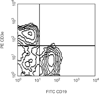
FITC Rat Anti-Mouse CD19 RUO
サイズ
0.5 mg
カタログ番号
553785
560591 Rev. 1

Please refer to Support Documents for Quality Certificates
Global - Refer to manufacturer's instructions for use and related User Manuals and Technical data sheets before using this products as described
Comparisons, where applicable, are made against older BD Technology, manual methods or are general performance claims. Comparisons are not made against non-BD technologies, unless otherwise noted.
For Research Use Only. Not for use in diagnostic or therapeutic procedures.