
-
抗体試薬
- フローサイトメトリー用試薬
-
ウェスタンブロッティング抗体試薬
- イムノアッセイ試薬
-
シングルセル試薬
- BD® AbSeq Assay
- BD Rhapsody™ Accessory Kits
- BD® OMICS-One Immune Profiler Protein Panel
- BD® Single-Cell Multiplexing Kit
- BD Rhapsody™ TCR/BCR Next Multiomic Assays
- BD Rhapsody™ Targeted mRNA Kits
- BD Rhapsody™ Whole Transcriptome Analysis (WTA) Amplification Kit
- BD® OMICS-Guard Sample Preservation Buffer
- BD Rhapsody™ ATAC-Seq Assays
- BD® OMICS-One Protein Panels
-
細胞機能評価のための試薬
-
顕微鏡・イメージング用試薬
-
細胞調製・分離試薬
-
- BD® AbSeq Assay
- BD Rhapsody™ Accessory Kits
- BD® OMICS-One Immune Profiler Protein Panel
- BD® Single-Cell Multiplexing Kit
- BD Rhapsody™ TCR/BCR Next Multiomic Assays
- BD Rhapsody™ Targeted mRNA Kits
- BD Rhapsody™ Whole Transcriptome Analysis (WTA) Amplification Kit
- BD® OMICS-Guard Sample Preservation Buffer
- BD Rhapsody™ ATAC-Seq Assays
- BD® OMICS-One Protein Panels
- Japan (Japanese)
-
Change country/language
Old Browser
Looks like you're visiting us from United States.
Would you like to stay on the current country site or be switched to your country?
BD Horizon™ BV421 Rat Anti-Mouse CD140b (PDGFRβ)
クローン APB5 (RUO)

Multicolor flow cytometric analysis of the correlated expression of CD140a and CD140b (PDGFRβ) on Mouse NIH/3T3 cells. Cells from the Mouse NIH/3T3 (Mouse embryonic fibroblast, ATCC® CRL-1658™) cell line were stained with BD Horizon™ BV421 Rat IgG2a, κ Isotype Control (Cat. No. 562602) and PE Rat IgG2a, κ Isotype Control (Cat. No. 553930) [Left Plot] or with BD Horizon™ BV421 Rat Anti-Mouse CD140b (PDGFRβ) antibody (Cat. No. 570045/570126) and PE Rat Anti-Mouse CD140a antibody (Cat. No. 562776) [Right Plot] at 0.25 µg/test. BD Via-Probe™ Cell Viability 7-AAD Solution (Cat. No. 555815/555816) was added to cells right before analysis. The bivariate pseudocolor density plot showing the correlated expression of CD140b (PDGFRβ) versus CD140a (or Ig Isotype control staining) was derived from gated events with the forward and side light-scatter characteristics of viable (7-AAD-negative) cells. Flow cytometry and data analysis were performed using a BD LSRFortessa™ X-20 Cell Analyzer System and FlowJo™ Software. Data shown on this Technical Data Sheet are not lot specific.

Flow cytometric analysis of CD140b (PDGFRβ) expression on Mouse splenic leukocytes and Mouse NIH/3T3 cells. C57BL/6 Mouse splenocytes (Left Plot) were preincubated with Purified Rat Anti-Mouse CD16/CD32 antibody (Mouse BD Fc Block™) [Cat. No. 553141/553142]. The splenic leukocytes and cells from the Mouse NIH/3T3 (Mouse embryonic fibroblast, ATCC® CRL-1658™) cell line (Right Plot) were then stained with either BD Horizon™ BV421 Rat IgG2a, κ Isotype Control (Cat. No. 562602; dashed line histograms) or BD Horizon™ BV421 Rat Anti-Mouse CD140b (PDGFRβ) antibody (Cat. No. 570045/570126; solid line histograms) at 0.25 µg/test. BD Via-Probe™ Cell Viability 7-AAD Solution (Cat. No. 555815/555816) was added to cells right before analysis. The fluorescence histograms showing CD140b (PDGFRβ) expression (or Ig Isotype control staining) were derived from gated events with the forward and side light-scatter characteristics of viable (7-AAD-negative) cells. Flow cytometry and data analysis were performed using a BD LSRFortessa™ X-20 Cell Analyzer System and FlowJo™ Software. Data shown on this Technical Data Sheet are not lot specific.


Multicolor flow cytometric analysis of the correlated expression of CD140a and CD140b (PDGFRβ) on Mouse NIH/3T3 cells. Cells from the Mouse NIH/3T3 (Mouse embryonic fibroblast, ATCC® CRL-1658™) cell line were stained with BD Horizon™ BV421 Rat IgG2a, κ Isotype Control (Cat. No. 562602) and PE Rat IgG2a, κ Isotype Control (Cat. No. 553930) [Left Plot] or with BD Horizon™ BV421 Rat Anti-Mouse CD140b (PDGFRβ) antibody (Cat. No. 570045/570126) and PE Rat Anti-Mouse CD140a antibody (Cat. No. 562776) [Right Plot] at 0.25 µg/test. BD Via-Probe™ Cell Viability 7-AAD Solution (Cat. No. 555815/555816) was added to cells right before analysis. The bivariate pseudocolor density plot showing the correlated expression of CD140b (PDGFRβ) versus CD140a (or Ig Isotype control staining) was derived from gated events with the forward and side light-scatter characteristics of viable (7-AAD-negative) cells. Flow cytometry and data analysis were performed using a BD LSRFortessa™ X-20 Cell Analyzer System and FlowJo™ Software. Data shown on this Technical Data Sheet are not lot specific.
Flow cytometric analysis of CD140b (PDGFRβ) expression on Mouse splenic leukocytes and Mouse NIH/3T3 cells. C57BL/6 Mouse splenocytes (Left Plot) were preincubated with Purified Rat Anti-Mouse CD16/CD32 antibody (Mouse BD Fc Block™) [Cat. No. 553141/553142]. The splenic leukocytes and cells from the Mouse NIH/3T3 (Mouse embryonic fibroblast, ATCC® CRL-1658™) cell line (Right Plot) were then stained with either BD Horizon™ BV421 Rat IgG2a, κ Isotype Control (Cat. No. 562602; dashed line histograms) or BD Horizon™ BV421 Rat Anti-Mouse CD140b (PDGFRβ) antibody (Cat. No. 570045/570126; solid line histograms) at 0.25 µg/test. BD Via-Probe™ Cell Viability 7-AAD Solution (Cat. No. 555815/555816) was added to cells right before analysis. The fluorescence histograms showing CD140b (PDGFRβ) expression (or Ig Isotype control staining) were derived from gated events with the forward and side light-scatter characteristics of viable (7-AAD-negative) cells. Flow cytometry and data analysis were performed using a BD LSRFortessa™ X-20 Cell Analyzer System and FlowJo™ Software. Data shown on this Technical Data Sheet are not lot specific.

Multicolor flow cytometric analysis of the correlated expression of CD140a and CD140b (PDGFRβ) on Mouse NIH/3T3 cells. Cells from the Mouse NIH/3T3 (Mouse embryonic fibroblast, ATCC® CRL-1658™) cell line were stained with BD Horizon™ BV421 Rat IgG2a, κ Isotype Control (Cat. No. 562602) and PE Rat IgG2a, κ Isotype Control (Cat. No. 553930) [Left Plot] or with BD Horizon™ BV421 Rat Anti-Mouse CD140b (PDGFRβ) antibody (Cat. No. 570045/570126) and PE Rat Anti-Mouse CD140a antibody (Cat. No. 562776) [Right Plot] at 0.25 µg/test. BD Via-Probe™ Cell Viability 7-AAD Solution (Cat. No. 555815/555816) was added to cells right before analysis. The bivariate pseudocolor density plot showing the correlated expression of CD140b (PDGFRβ) versus CD140a (or Ig Isotype control staining) was derived from gated events with the forward and side light-scatter characteristics of viable (7-AAD-negative) cells. Flow cytometry and data analysis were performed using a BD LSRFortessa™ X-20 Cell Analyzer System and FlowJo™ Software. Data shown on this Technical Data Sheet are not lot specific.

Flow cytometric analysis of CD140b (PDGFRβ) expression on Mouse splenic leukocytes and Mouse NIH/3T3 cells. C57BL/6 Mouse splenocytes (Left Plot) were preincubated with Purified Rat Anti-Mouse CD16/CD32 antibody (Mouse BD Fc Block™) [Cat. No. 553141/553142]. The splenic leukocytes and cells from the Mouse NIH/3T3 (Mouse embryonic fibroblast, ATCC® CRL-1658™) cell line (Right Plot) were then stained with either BD Horizon™ BV421 Rat IgG2a, κ Isotype Control (Cat. No. 562602; dashed line histograms) or BD Horizon™ BV421 Rat Anti-Mouse CD140b (PDGFRβ) antibody (Cat. No. 570045/570126; solid line histograms) at 0.25 µg/test. BD Via-Probe™ Cell Viability 7-AAD Solution (Cat. No. 555815/555816) was added to cells right before analysis. The fluorescence histograms showing CD140b (PDGFRβ) expression (or Ig Isotype control staining) were derived from gated events with the forward and side light-scatter characteristics of viable (7-AAD-negative) cells. Flow cytometry and data analysis were performed using a BD LSRFortessa™ X-20 Cell Analyzer System and FlowJo™ Software. Data shown on this Technical Data Sheet are not lot specific.


BD Horizon™ BV421 Rat Anti-Mouse CD140b (PDGFRβ)

BD Horizon™ BV421 Rat Anti-Mouse CD140b (PDGFRβ)

Regulatory Statusの凡例
Any use of products other than the permitted use without the express written authorization of Becton, Dickinson and Company is strictly prohibited.
Preparation and Storage
推奨アッセイ手順
BD® CompBeads can be used as surrogates to assess fluorescence spillover (compensation). When fluorochrome conjugated antibodies are bound to BD® CompBeads, they have spectral properties very similar to cells. However, for some fluorochromes there can be small differences in spectral emissions compared to cells, resulting in spillover values that differ when compared to biological controls. It is strongly recommended that when using a reagent for the first time, users compare the spillover on cells and BD® CompBeads to ensure that BD® CompBeads are appropriate for your specific cellular application.
For optimal and reproducible results, BD Horizon Brilliant Stain Buffer should be used anytime BD Horizon Brilliant dyes are used in a multicolor flow cytometry panel. Fluorescent dye interactions may cause staining artifacts which may affect data interpretation. The BD Horizon Brilliant Stain Buffer was designed to minimize these interactions. When BD Horizon Brilliant Stain Buffer is used in in the multicolor panel, it should also be used in the corresponding compensation controls for all dyes to achieve the most accurate compensation. For the most accurate compensation, compensation controls created with either cells or beads should be exposed to BD Horizon Brilliant Stain Buffer for the same length of time as the corresponding multicolor panel. More information can be found in the Technical Data Sheet of the BD Horizon Brilliant Stain Buffer (Cat. No. 563794/566349) or the BD Horizon Brilliant Stain Buffer Plus (Cat. No. 566385).
Product Notices
- Please refer to www.bdbiosciences.com/us/s/resources for technical protocols.
- Since applications vary, each investigator should titrate the reagent to obtain optimal results.
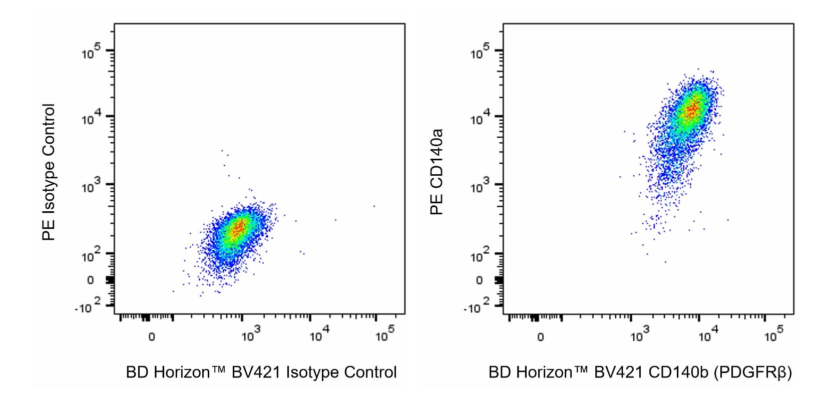
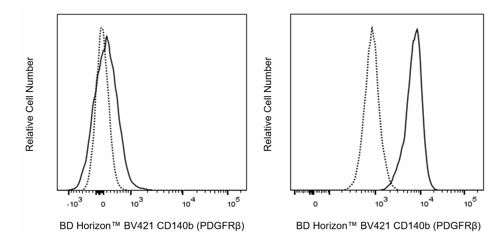
- An isotype control should be used at the same concentration as the antibody of interest.
- Caution: Sodium azide yields highly toxic hydrazoic acid under acidic conditions. Dilute azide compounds in running water before discarding to avoid accumulation of potentially explosive deposits in plumbing.
- For fluorochrome spectra and suitable instrument settings, please refer to our Multicolor Flow Cytometry web page at www.bdbiosciences.com/colors.
- Please refer to http://regdocs.bd.com to access safety data sheets (SDS).
- For U.S. patents that may apply, see bd.com/patents.
関連製品


The APB5 monoclonal antibody specifically recognizes CD140b, the Platelet-Derived Growth Factor Receptor beta chain (PDGFRβ). CD140b is a single-pass type I transmembrane glycoprotein that contains an intracellular tyrosine kinase domain. This receptor is encoded by Pdgfrb and is widely expressed on cells within embryonic tissues and cells of mesenchymal origin in the adult mouse including fibroblasts and vascular smooth muscle cells. CD140b forms homodimers or heterodimeric receptor complexes with CD140a (PDGFRα) upon binding various dimeric Platelet-Derived Growth Factors (PDGF) isoforms that lead to receptor activation. Upon activation, CD140b is autophosphorylated at multiple tyrosine sites and, in turn, initiates several signaling cascades by binding and activation of cytoplasmic SH2 domain-containing signal transduction molecules, such as GRB2, Src, GAP, PI3 kinase, PLCγ, and NCK. These signaling pathways regulate cellular growth and proliferation, survival, differentiation, and migration involved in the development of embryonic tissues, blood vessel formation, wound healing, and in a range of diseases including fibrotic conditions, atherosclerosis, and malignancies.

Development References (7)
-
Andrae J, Gallini R, Betsholtz C. Role of platelet-derived growth factors in physiology and medicine.. Genes Dev. 2008; 22(10):1276-312. (Biology). View Reference
-
Cossarizza A, Chang HD, Radbruch A, et al. Guidelines for the use of flow cytometry and cell sorting in immunological studies (second edition).. Eur J Immunol. 2019; 49(10):1457-1973. (Clone-specific). View Reference
-
Cremasco V, Woodruff MC, Onder L, et al. B cell homeostasis and follicle confines are governed by fibroblastic reticular cells.. Nat Immunol. 2014; 15(10):973-81. (Clone-specific: Flow cytometry). View Reference
-
Ostman A, Heldin CH. Involvement of platelet-derived growth factor in disease: development of specific antagonists.. Adv Cancer Res. 2001; 80:1-38. (Biology). View Reference
-
Patenaude J, Perreault C. Thymic Mesenchymal Cells Have a Distinct Transcriptomic Profile.. J Immunol. 2016; 196(11):4760-70. (Clone-specific: Flow cytometry). View Reference
-
Sano H, Sudo T, Yokode M, et al. Functional blockade of platelet-derived growth factor receptor-beta but not of receptor-alpha prevents vascular smooth muscle cell accumulation in fibrous cap lesions in apolipoprotein E-deficient mice.. Circulation. 2001; 103(24):2955-60. (Immunogen: Functional assay, Immunohistochemistry, Western blot). View Reference
-
Sano H, Yokode M, Takakura N, et al. Study on PDGF receptor beta pathway in glomerular formation in neonate mice.. Ann N Y Acad Sci. 2001; 947:303-5. (Clone-specific: Immunohistochemistry, Western blot). View Reference
Please refer to Support Documents for Quality Certificates
Global - Refer to manufacturer's instructions for use and related User Manuals and Technical data sheets before using this products as described
Comparisons, where applicable, are made against older BD Technology, manual methods or are general performance claims. Comparisons are not made against non-BD technologies, unless otherwise noted.
For Research Use Only. Not for use in diagnostic or therapeutic procedures.