
-
抗体試薬
- フローサイトメトリー用試薬
-
ウェスタンブロッティング抗体試薬
- イムノアッセイ試薬
-
シングルセル試薬
- BD® AbSeq Assay
- BD Rhapsody™ Accessory Kits
- BD® OMICS-One Immune Profiler Protein Panel
- BD® Single-Cell Multiplexing Kit
- BD Rhapsody™ TCR/BCR Next Multiomic Assays
- BD Rhapsody™ Targeted mRNA Kits
- BD Rhapsody™ Whole Transcriptome Analysis (WTA) Amplification Kit
- BD® OMICS-Guard Sample Preservation Buffer
- BD Rhapsody™ ATAC-Seq Assays
- BD® OMICS-One Protein Panels
-
細胞機能評価のための試薬
-
顕微鏡・イメージング用試薬
-
細胞調製・分離試薬
-
- BD® AbSeq Assay
- BD Rhapsody™ Accessory Kits
- BD® OMICS-One Immune Profiler Protein Panel
- BD® Single-Cell Multiplexing Kit
- BD Rhapsody™ TCR/BCR Next Multiomic Assays
- BD Rhapsody™ Targeted mRNA Kits
- BD Rhapsody™ Whole Transcriptome Analysis (WTA) Amplification Kit
- BD® OMICS-Guard Sample Preservation Buffer
- BD Rhapsody™ ATAC-Seq Assays
- BD® OMICS-One Protein Panels
- Japan (Japanese)
-
Change country/language
Old Browser
Looks like you're visiting us from United States.
Would you like to stay on the current country site or be switched to your country?
BD Horizon™ R718 Mouse Anti-Mouse CD369 (CLEC7A)
クローン 2F3 (RUO)

Multicolor flow cytometric analysis of CD369 (CLEC7A) expression on viable Mouse peritoneal exudate cells (PEC). BALB/c Mouse thioglycolate-elicited PEC cells were harvested and pre-incubated with Purified Rat Anti-Mouse CD16/CD32 antibody (Mouse BD Fc Block™) [Cat. No. 553141/553142]. The cells were then stained with PE Rat Anti-Mouse F4/80 antibody (Cat. No. 565410) and with either BD Horizon™ R718 Mouse IgG1, κ Isotype Control (Cat. No. 567470; Left Plot) or BD Horizon™ R718 Mouse Anti-Mouse CD369 (CLEC7A) antibody (Cat. No. 569951/569956; Right Plot) at 1 µg/test. BD Via-Probe™ Cell Viability 7-AAD Solution was added to cells right before analysis. The bivariate pseudocolor density plot showing the correlated expression of CD369 (CLEC7A) [or Ig Isotype control staining] versus F4/80 was derived from 7-AAD-negative gated events with the forward and side light-scatter characteristics of viable PEC cells. Flow cytometry and data analysis were performed using a BD LSRFortessa™ X-20 Cell Analyzer System and FlowJo™ software. Data shown on this Technical Data Sheet are not lot specific.

Multicolor flow cytometric analysis of CD369 (CLEC7A) expression on Mouse peripheral blood leukocytes (PBL). BALB/c Mouse peripheral blood leukocytes (PBL) were stained with PE Rat Anti-Mouse Ly-6G antibody (Cat. No. 551461) and with either BD Horizon™ R718 Mouse IgG1, κ Isotype Control (Cat. No. 567470; Left Plot) or BD Horizon™ R718 Mouse Anti-Mouse CD369 (CLEC7A) antibody (Cat. No. 569951/569956; Right Plot) at 1 µg/test. Erythrocytes were lysed with BD Pharm Lyse™ Lysing Buffer (Cat. No. 555899). The bivariate pseudocolor density plot showing the correlated expression of CD369 (CLEC7A) [or Ig Isotype control staining] versus Ly-6G was derived from gated events with the forward and side light-scatter characteristics of viable leukocyte populations. Flow cytometry and data analysis were performed using a BD LSRFortessa™ X-20 Cell Analyzer System and FlowJo™ software. Data shown on this Technical Data Sheet are not lot specific.


Multicolor flow cytometric analysis of CD369 (CLEC7A) expression on viable Mouse peritoneal exudate cells (PEC). BALB/c Mouse thioglycolate-elicited PEC cells were harvested and pre-incubated with Purified Rat Anti-Mouse CD16/CD32 antibody (Mouse BD Fc Block™) [Cat. No. 553141/553142]. The cells were then stained with PE Rat Anti-Mouse F4/80 antibody (Cat. No. 565410) and with either BD Horizon™ R718 Mouse IgG1, κ Isotype Control (Cat. No. 567470; Left Plot) or BD Horizon™ R718 Mouse Anti-Mouse CD369 (CLEC7A) antibody (Cat. No. 569951/569956; Right Plot) at 1 µg/test. BD Via-Probe™ Cell Viability 7-AAD Solution was added to cells right before analysis. The bivariate pseudocolor density plot showing the correlated expression of CD369 (CLEC7A) [or Ig Isotype control staining] versus F4/80 was derived from 7-AAD-negative gated events with the forward and side light-scatter characteristics of viable PEC cells. Flow cytometry and data analysis were performed using a BD LSRFortessa™ X-20 Cell Analyzer System and FlowJo™ software. Data shown on this Technical Data Sheet are not lot specific.
Multicolor flow cytometric analysis of CD369 (CLEC7A) expression on Mouse peripheral blood leukocytes (PBL). BALB/c Mouse peripheral blood leukocytes (PBL) were stained with PE Rat Anti-Mouse Ly-6G antibody (Cat. No. 551461) and with either BD Horizon™ R718 Mouse IgG1, κ Isotype Control (Cat. No. 567470; Left Plot) or BD Horizon™ R718 Mouse Anti-Mouse CD369 (CLEC7A) antibody (Cat. No. 569951/569956; Right Plot) at 1 µg/test. Erythrocytes were lysed with BD Pharm Lyse™ Lysing Buffer (Cat. No. 555899). The bivariate pseudocolor density plot showing the correlated expression of CD369 (CLEC7A) [or Ig Isotype control staining] versus Ly-6G was derived from gated events with the forward and side light-scatter characteristics of viable leukocyte populations. Flow cytometry and data analysis were performed using a BD LSRFortessa™ X-20 Cell Analyzer System and FlowJo™ software. Data shown on this Technical Data Sheet are not lot specific.

Multicolor flow cytometric analysis of CD369 (CLEC7A) expression on viable Mouse peritoneal exudate cells (PEC). BALB/c Mouse thioglycolate-elicited PEC cells were harvested and pre-incubated with Purified Rat Anti-Mouse CD16/CD32 antibody (Mouse BD Fc Block™) [Cat. No. 553141/553142]. The cells were then stained with PE Rat Anti-Mouse F4/80 antibody (Cat. No. 565410) and with either BD Horizon™ R718 Mouse IgG1, κ Isotype Control (Cat. No. 567470; Left Plot) or BD Horizon™ R718 Mouse Anti-Mouse CD369 (CLEC7A) antibody (Cat. No. 569951/569956; Right Plot) at 1 µg/test. BD Via-Probe™ Cell Viability 7-AAD Solution was added to cells right before analysis. The bivariate pseudocolor density plot showing the correlated expression of CD369 (CLEC7A) [or Ig Isotype control staining] versus F4/80 was derived from 7-AAD-negative gated events with the forward and side light-scatter characteristics of viable PEC cells. Flow cytometry and data analysis were performed using a BD LSRFortessa™ X-20 Cell Analyzer System and FlowJo™ software. Data shown on this Technical Data Sheet are not lot specific.

Multicolor flow cytometric analysis of CD369 (CLEC7A) expression on Mouse peripheral blood leukocytes (PBL). BALB/c Mouse peripheral blood leukocytes (PBL) were stained with PE Rat Anti-Mouse Ly-6G antibody (Cat. No. 551461) and with either BD Horizon™ R718 Mouse IgG1, κ Isotype Control (Cat. No. 567470; Left Plot) or BD Horizon™ R718 Mouse Anti-Mouse CD369 (CLEC7A) antibody (Cat. No. 569951/569956; Right Plot) at 1 µg/test. Erythrocytes were lysed with BD Pharm Lyse™ Lysing Buffer (Cat. No. 555899). The bivariate pseudocolor density plot showing the correlated expression of CD369 (CLEC7A) [or Ig Isotype control staining] versus Ly-6G was derived from gated events with the forward and side light-scatter characteristics of viable leukocyte populations. Flow cytometry and data analysis were performed using a BD LSRFortessa™ X-20 Cell Analyzer System and FlowJo™ software. Data shown on this Technical Data Sheet are not lot specific.

BD Horizon™ R718 Mouse Anti-Mouse CD369 (CLEC7A)

BD Horizon™ R718 Mouse Anti-Mouse CD369 (CLEC7A)
Regulatory Statusの凡例
Any use of products other than the permitted use without the express written authorization of Becton, Dickinson and Company is strictly prohibited.
Preparation and Storage
推奨アッセイ手順
BD® CompBeads can be used as surrogates to assess fluorescence spillover (compensation). When fluorochrome conjugated antibodies are bound to BD® CompBeads, they have spectral properties very similar to cells. However, for some fluorochromes there can be small differences in spectral emissions compared to cells, resulting in spillover values that differ when compared to biological controls. It is strongly recommended that when using a reagent for the first time, users compare the spillover on cells and BD® CompBeads to ensure that BD® CompBeads are appropriate for your specific cellular application.
Product Notices
- Please refer to www.bdbiosciences.com/us/s/resources for technical protocols.
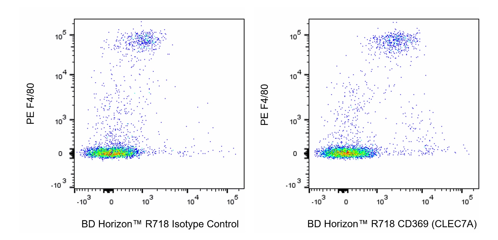
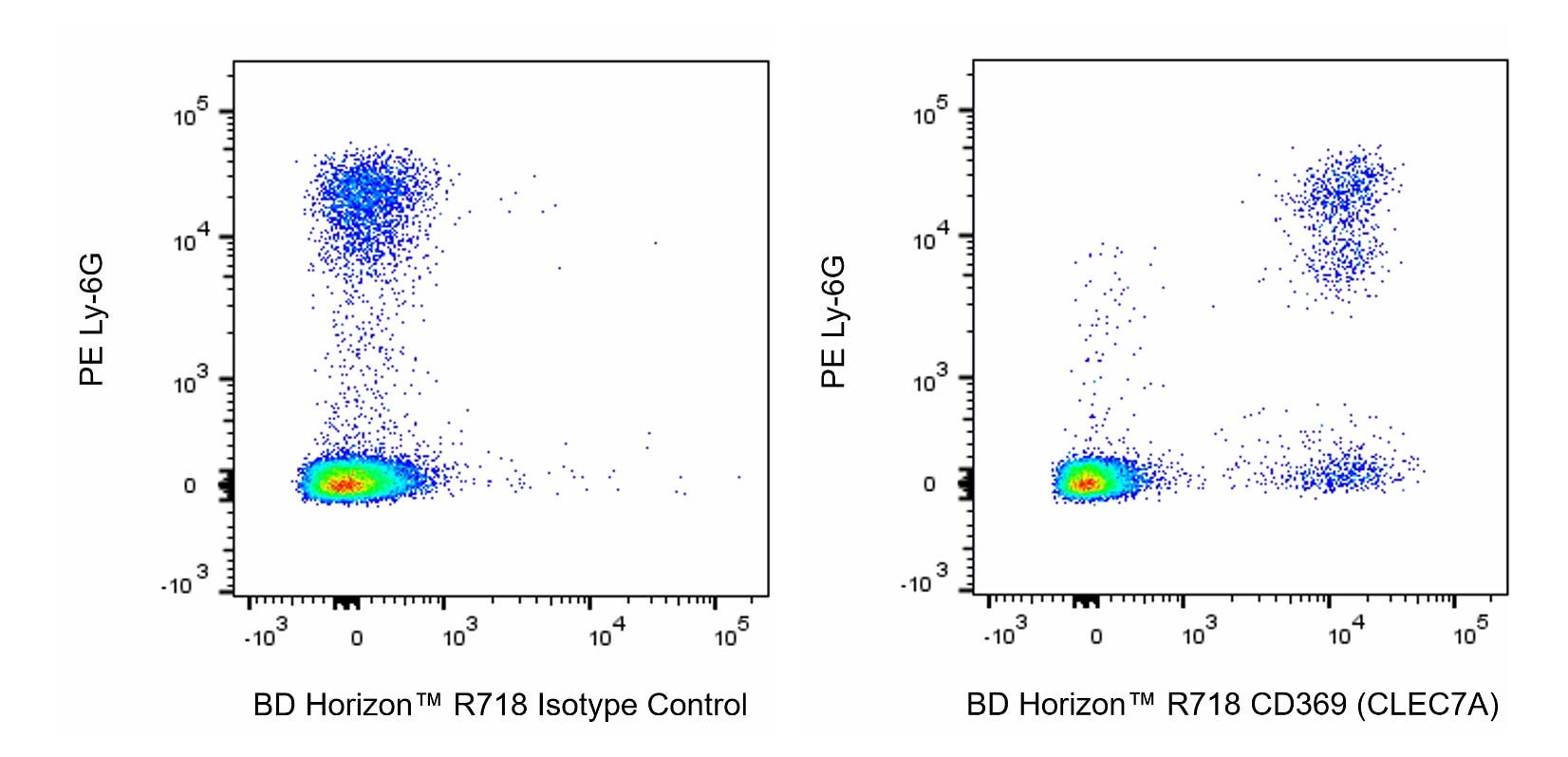
- Since applications vary, each investigator should titrate the reagent to obtain optimal results.
- An isotype control should be used at the same concentration as the antibody of interest.
- Caution: Sodium azide yields highly toxic hydrazoic acid under acidic conditions. Dilute azide compounds in running water before discarding to avoid accumulation of potentially explosive deposits in plumbing.
- For fluorochrome spectra and suitable instrument settings, please refer to our Multicolor Flow Cytometry web page at www.bdbiosciences.com/colors.
- This product is provided under an Agreement between BIOTIUM and BD Biosciences. This product, and only in the amount purchased by buyer, may be used solely for buyer’s own internal research, in a manner consistent with the accompanying product literature. No other right to use, sell or otherwise transfer (a) this product, or (b) its components is hereby granted expressly, by implication or by estoppel. This product is for research use only. Diagnostic uses require a separate license from Biotium, Inc. For information on purchasing a license to this product including for purposes other than research, contact Biotium, Inc., 3159 Corporate Place, Hayward, CA 94545, Tel: (510) 265-1027. Fax: (510) 265-1352. Email: btinfo@biotium.com.
- Please refer to http://regdocs.bd.com to access safety data sheets (SDS).
- Alexa Fluor™ is a trademark of Life Technologies Corporation.
- For U.S. patents that may apply, see bd.com/patents.
関連製品


最近閲覧済み
The 2F3 monoclonal antibody specifically recognizes CD369 which is also known as Dectin-1 (Dendritic cell-associated C-type lectin 1) or BGR (Beta-glucan receptor). CD369 is a ~ 43 kDa type II transmembrane C-type lectin that is encoded by Clec7a (C-type lectin domain family 7, member a). The extracellular portion of CD369 (Clec7a) contains a C-terminal stalk with a carbohydrate recognition domain (CRD) that is followed by a transmembrane segment, and an ITAM-containing cytoplasmic tail. This protein is predominantly expressed on monocytes/macrophages, neutrophils and some dendritic cell populations and at a lower level on a sub-population of T cells. CD369 (Clec7a) binds to beta-glucan polymers and functions as a pattern recognition receptor (PRR) in innate immune response to fungal and bacterial pathogens. CD369 mediated signaling may play a role in leucocyte responses, including phagocytosis or enhanced cytokine production. In mice, two functionally different full-length and stalkless isoforms have been characterized. Both isoforms vary in their ability to recognize zymosan and mediate cellular responses upon zymosan recognition.

Development References (4)
-
Brown GD, Taylor PR, Reid DM, et al. Dectin-1 is a major beta-glucan receptor on macrophages.. J Exp Med. 2002; 196(3):407-12. (Biology). View Reference
-
Herre J, Gordon S, Brown GD. Dectin-1 and its role in the recognition of beta-glucans by macrophages.. Mol Immunol. 2004; 40(12):869-76. (Biology). View Reference
-
Taylor PR, Brown GD, Reid DM, et al. The beta-glucan receptor, dectin-1, is predominantly expressed on the surface of cells of the monocyte/macrophage and neutrophil lineages.. J Immunol. 2002; 169(7):3876-82. (Biology). View Reference
-
Underhill DM, Rossnagle E, Lowell CA, Simmons RM. Dectin-1 activates Syk tyrosine kinase in a dynamic subset of macrophages for reactive oxygen production.. Blood. 2005; 106(7):2543-50. (Biology). View Reference
Please refer to Support Documents for Quality Certificates
Global - Refer to manufacturer's instructions for use and related User Manuals and Technical data sheets before using this products as described
Comparisons, where applicable, are made against older BD Technology, manual methods or are general performance claims. Comparisons are not made against non-BD technologies, unless otherwise noted.
For Research Use Only. Not for use in diagnostic or therapeutic procedures.