-
抗体試薬
- フローサイトメトリー用試薬
-
ウェスタンブロッティング抗体試薬
- イムノアッセイ試薬
-
シングルセル試薬
- BD® AbSeq Assay
- BD Rhapsody™ Accessory Kits
- BD® OMICS-One Immune Profiler Protein Panel
- BD® Single-Cell Multiplexing Kit
- BD Rhapsody™ TCR/BCR Next Multiomic Assays
- BD Rhapsody™ Targeted mRNA Kits
- BD Rhapsody™ Whole Transcriptome Analysis (WTA) Amplification Kit
- BD® OMICS-Guard Sample Preservation Buffer
- BD Rhapsody™ ATAC-Seq Assays
- BD® OMICS-One Protein Panels
-
細胞機能評価のための試薬
-
顕微鏡・イメージング用試薬
-
細胞調製・分離試薬
-
- BD® AbSeq Assay
- BD Rhapsody™ Accessory Kits
- BD® OMICS-One Immune Profiler Protein Panel
- BD® Single-Cell Multiplexing Kit
- BD Rhapsody™ TCR/BCR Next Multiomic Assays
- BD Rhapsody™ Targeted mRNA Kits
- BD Rhapsody™ Whole Transcriptome Analysis (WTA) Amplification Kit
- BD® OMICS-Guard Sample Preservation Buffer
- BD Rhapsody™ ATAC-Seq Assays
- BD® OMICS-One Protein Panels
- Japan (Japanese)
-
Change country/language
Old Browser
Looks like you're visiting us from United States.
Would you like to stay on the current country site or be switched to your country?
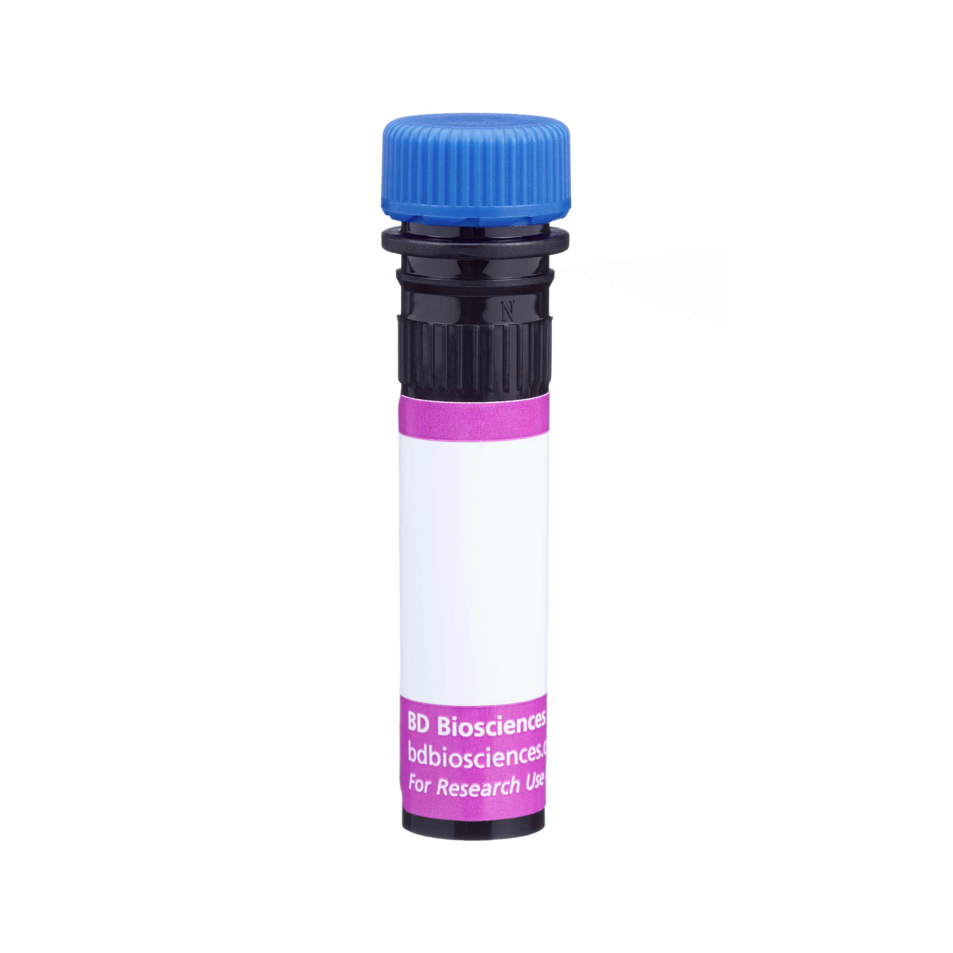
BD Horizon™ BV421 Rat Anti-Mouse Granzyme C
クローン SFC1D8.rMAb (RUO)

Multicolor flow cytometric analysis of Granzyme C expression in activated Mouse splenic leukocytes. C57BL/6 Mouse splenocytes were activated by culture with Recombinant Mouse IL-15 protein (100 ng/ml) for 4 days (37°C). The cells were harvested, washed, preincubated with Purified Rat Anti-Mouse CD16/CD32 antibody (Mouse BD Fc Block™) [Cat. No. 553141/553142] and then stained with APC Mouse Anti-Mouse NK-1.1 antibody (Cat. No. 550627) and BD Horizon™ Fixable Viability Stain 780 (Cat. No. 565388). The cells were then fixed with BD Cytofix™ Fixation Buffer (Cat. No. 554655), washed, permeabilized with and stained in BD Perm/Wash™ Buffer (Cat. No. 554723) with either BD Horizon™ BV421 Rat IgG1, κ Isotype Control (Cat. No. 562868; Left Plot) or BD Horizon™ BV421 Rat Anti-Mouse Granzyme C antibody (Cat. No. 569861/569937; Right Plot) at 0.5 µg/test. The bivariate pseudocolor density plot showing the correlated expression of Granzyme C (or Ig Isotype control staining) versus NK-1.1 was derived from gated events with the forward and side light-scatter characteristics of viable (Fixable Viability Stain 780-negative) splenocytes. Flow cytometry and data analysis were performed using a BD LSRFortessa™ X-20 Cell Analyzer System and FlowJo™ software. Data shown on this Technical Data Sheet are not lot specific.

Multicolor flow cytometric analysis of Granzyme C expression in activated Mouse splenic leukocytes. C57BL/6 Mouse splenocytes were activated by culture with Recombinant Mouse IL-15 protein (100 ng/ml) for 4 days (37°C). The cells were harvested, washed, preincubated with Purified Rat Anti-Mouse CD16/CD32 antibody (Mouse BD Fc Block™) [Cat. No. 553141/553142] and then stained with APC Mouse Anti-Mouse NK-1.1 antibody (Cat. No. 550627) and BD Horizon™ Fixable Viability Stain 780 (Cat. No. 565388). The cells were then fixed with BD Cytofix™ Fixation Buffer (Cat. No. 554655), washed, permeabilized with and stained in BD Perm/Wash™ Buffer (Cat. No. 554723) with either BD Horizon™ BV421 Rat IgG1, κ Isotype Control (Cat. No. 562868; Left Plot) or BD Horizon™ BV421 Rat Anti-Mouse Granzyme C antibody (Cat. No. 569861/569937; Right Plot) at 0.5 µg/test. The bivariate pseudocolor density plot showing the correlated expression of Granzyme C (or Ig Isotype control staining) versus NK-1.1 was derived from gated events with the forward and side light-scatter characteristics of viable (Fixable Viability Stain 780-negative) splenocytes. Flow cytometry and data analysis were performed using a BD LSRFortessa™ X-20 Cell Analyzer System and FlowJo™ software. Data shown on this Technical Data Sheet are not lot specific.


Multicolor flow cytometric analysis of Granzyme C expression in activated Mouse splenic leukocytes. C57BL/6 Mouse splenocytes were activated by culture with Recombinant Mouse IL-15 protein (100 ng/ml) for 4 days (37°C). The cells were harvested, washed, preincubated with Purified Rat Anti-Mouse CD16/CD32 antibody (Mouse BD Fc Block™) [Cat. No. 553141/553142] and then stained with APC Mouse Anti-Mouse NK-1.1 antibody (Cat. No. 550627) and BD Horizon™ Fixable Viability Stain 780 (Cat. No. 565388). The cells were then fixed with BD Cytofix™ Fixation Buffer (Cat. No. 554655), washed, permeabilized with and stained in BD Perm/Wash™ Buffer (Cat. No. 554723) with either BD Horizon™ BV421 Rat IgG1, κ Isotype Control (Cat. No. 562868; Left Plot) or BD Horizon™ BV421 Rat Anti-Mouse Granzyme C antibody (Cat. No. 569861/569937; Right Plot) at 0.5 µg/test. The bivariate pseudocolor density plot showing the correlated expression of Granzyme C (or Ig Isotype control staining) versus NK-1.1 was derived from gated events with the forward and side light-scatter characteristics of viable (Fixable Viability Stain 780-negative) splenocytes. Flow cytometry and data analysis were performed using a BD LSRFortessa™ X-20 Cell Analyzer System and FlowJo™ software. Data shown on this Technical Data Sheet are not lot specific.
Multicolor flow cytometric analysis of Granzyme C expression in activated Mouse splenic leukocytes. C57BL/6 Mouse splenocytes were activated by culture with Recombinant Mouse IL-15 protein (100 ng/ml) for 4 days (37°C). The cells were harvested, washed, preincubated with Purified Rat Anti-Mouse CD16/CD32 antibody (Mouse BD Fc Block™) [Cat. No. 553141/553142] and then stained with APC Mouse Anti-Mouse NK-1.1 antibody (Cat. No. 550627) and BD Horizon™ Fixable Viability Stain 780 (Cat. No. 565388). The cells were then fixed with BD Cytofix™ Fixation Buffer (Cat. No. 554655), washed, permeabilized with and stained in BD Perm/Wash™ Buffer (Cat. No. 554723) with either BD Horizon™ BV421 Rat IgG1, κ Isotype Control (Cat. No. 562868; Left Plot) or BD Horizon™ BV421 Rat Anti-Mouse Granzyme C antibody (Cat. No. 569861/569937; Right Plot) at 0.5 µg/test. The bivariate pseudocolor density plot showing the correlated expression of Granzyme C (or Ig Isotype control staining) versus NK-1.1 was derived from gated events with the forward and side light-scatter characteristics of viable (Fixable Viability Stain 780-negative) splenocytes. Flow cytometry and data analysis were performed using a BD LSRFortessa™ X-20 Cell Analyzer System and FlowJo™ software. Data shown on this Technical Data Sheet are not lot specific.

Multicolor flow cytometric analysis of Granzyme C expression in activated Mouse splenic leukocytes. C57BL/6 Mouse splenocytes were activated by culture with Recombinant Mouse IL-15 protein (100 ng/ml) for 4 days (37°C). The cells were harvested, washed, preincubated with Purified Rat Anti-Mouse CD16/CD32 antibody (Mouse BD Fc Block™) [Cat. No. 553141/553142] and then stained with APC Mouse Anti-Mouse NK-1.1 antibody (Cat. No. 550627) and BD Horizon™ Fixable Viability Stain 780 (Cat. No. 565388). The cells were then fixed with BD Cytofix™ Fixation Buffer (Cat. No. 554655), washed, permeabilized with and stained in BD Perm/Wash™ Buffer (Cat. No. 554723) with either BD Horizon™ BV421 Rat IgG1, κ Isotype Control (Cat. No. 562868; Left Plot) or BD Horizon™ BV421 Rat Anti-Mouse Granzyme C antibody (Cat. No. 569861/569937; Right Plot) at 0.5 µg/test. The bivariate pseudocolor density plot showing the correlated expression of Granzyme C (or Ig Isotype control staining) versus NK-1.1 was derived from gated events with the forward and side light-scatter characteristics of viable (Fixable Viability Stain 780-negative) splenocytes. Flow cytometry and data analysis were performed using a BD LSRFortessa™ X-20 Cell Analyzer System and FlowJo™ software. Data shown on this Technical Data Sheet are not lot specific.

Multicolor flow cytometric analysis of Granzyme C expression in activated Mouse splenic leukocytes. C57BL/6 Mouse splenocytes were activated by culture with Recombinant Mouse IL-15 protein (100 ng/ml) for 4 days (37°C). The cells were harvested, washed, preincubated with Purified Rat Anti-Mouse CD16/CD32 antibody (Mouse BD Fc Block™) [Cat. No. 553141/553142] and then stained with APC Mouse Anti-Mouse NK-1.1 antibody (Cat. No. 550627) and BD Horizon™ Fixable Viability Stain 780 (Cat. No. 565388). The cells were then fixed with BD Cytofix™ Fixation Buffer (Cat. No. 554655), washed, permeabilized with and stained in BD Perm/Wash™ Buffer (Cat. No. 554723) with either BD Horizon™ BV421 Rat IgG1, κ Isotype Control (Cat. No. 562868; Left Plot) or BD Horizon™ BV421 Rat Anti-Mouse Granzyme C antibody (Cat. No. 569861/569937; Right Plot) at 0.5 µg/test. The bivariate pseudocolor density plot showing the correlated expression of Granzyme C (or Ig Isotype control staining) versus NK-1.1 was derived from gated events with the forward and side light-scatter characteristics of viable (Fixable Viability Stain 780-negative) splenocytes. Flow cytometry and data analysis were performed using a BD LSRFortessa™ X-20 Cell Analyzer System and FlowJo™ software. Data shown on this Technical Data Sheet are not lot specific.


Regulatory Statusの凡例
Any use of products other than the permitted use without the express written authorization of Becton, Dickinson and Company is strictly prohibited.
Preparation and Storage
推奨アッセイ手順
BD® CompBeads can be used as surrogates to assess fluorescence spillover (compensation). When fluorochrome conjugated antibodies are bound to BD® CompBeads, they have spectral properties very similar to cells. However, for some fluorochromes there can be small differences in spectral emissions compared to cells, resulting in spillover values that differ when compared to biological controls. It is strongly recommended that when using a reagent for the first time, users compare the spillover on cells and BD® CompBeads to ensure that BD® CompBeads are appropriate for your specific cellular application.
For optimal and reproducible results, BD Horizon Brilliant Stain Buffer should be used anytime BD Horizon Brilliant dyes are used in a multicolor flow cytometry panel. Fluorescent dye interactions may cause staining artifacts which may affect data interpretation. The BD Horizon Brilliant Stain Buffer was designed to minimize these interactions. When BD Horizon Brilliant Stain Buffer is used in in the multicolor panel, it should also be used in the corresponding compensation controls for all dyes to achieve the most accurate compensation. For the most accurate compensation, compensation controls created with either cells or beads should be exposed to BD Horizon Brilliant Stain Buffer for the same length of time as the corresponding multicolor panel. More information can be found in the Technical Data Sheet of the BD Horizon Brilliant Stain Buffer (Cat. No. 563794/566349) or the BD Horizon Brilliant Stain Buffer Plus (Cat. No. 566385).
Product Notices
- Please refer to www.bdbiosciences.com/us/s/resources for technical protocols.
- Since applications vary, each investigator should titrate the reagent to obtain optimal results.
- An isotype control should be used at the same concentration as the antibody of interest.
- Caution: Sodium azide yields highly toxic hydrazoic acid under acidic conditions. Dilute azide compounds in running water before discarding to avoid accumulation of potentially explosive deposits in plumbing.
- For fluorochrome spectra and suitable instrument settings, please refer to our Multicolor Flow Cytometry web page at www.bdbiosciences.com/colors.
- Please refer to http://regdocs.bd.com to access safety data sheets (SDS).
- For U.S. patents that may apply, see bd.com/patents.
関連製品


The SFC1D8.rMAb is a recombinant Rat IgG1 κ monoclonal antibody with VH and VL regions derived from SFC1D8 hybridoma cells that secrete Armenian Hamster IgG antibodies specific for Mouse Granzyme C. Granzyme C is also known as Cytotoxic cell protease 2 (CCP2) or Ctla5 (Ctla-5). Granzyme C is a serine protease that is encoded by Gzmc (granzyme C) which belongs to the peptidase S1 family. Granzyme C is found in the cytotoxic granules of cytotoxic T lymphocyte and natural killer (NK) effector cells. Upon recognition of target cells, these effector cells can exocytose Granzyme C which induces apoptosis of target cells leading to their externalization of phosphatidylserine, nuclear condensation, mitochondrial swelling, and the single-stranded DNA nicks. Granzyme C can reportedly support cytotoxic T lymphocyte-mediated killing of target cells in the absence of Granzyme A or B.
Development References (9)
-
Cai SF, Fehniger TA, Cao X, et al. Differential expression of granzyme B and C in murine cytotoxic lymphocytes.. J Immunol. 2009; 182(10):6287-97. (Immunogen: ELISA, Intracellular Staining/Flow Cytometry). View Reference
-
Cao X, Cai SF, Fehniger TA, et al. Granzyme B and perforin are important for regulatory T cell-mediated suppression of tumor clearance.. Immunity. 2007; 27(4):635-46. (Biology). View Reference
-
Garcia-Sanz JA, MacDonald HR, Jenne DE, Tschopp J, Nabholz M. Cell specificity of granzyme gene expression.. J Immunol. 1990; 145(9):3111-8. (Biology). View Reference
-
Getachew Y, Stout-Delgado H, Miller BC, Thiele DL. Granzyme C supports efficient CTL-mediated killing late in primary alloimmune responses.. J Immunol. 2008; 181(11):7810-7. (Biology). View Reference
-
Janas ML, Groves P, Kienzle N, Kelso A. IL-2 regulates perforin and granzyme gene expression in CD8+ T cells independently of its effects on survival and proliferation.. J Immunol. 2005; 175(12):8003-10. (Biology). View Reference
-
Jenne D, Rey C, Masson D, et al. cDNA cloning of granzyme C, a granule-associated serine protease of cytolytic T lymphocytes.. J Immunol. 1988; 140(1):318-23. (Biology). View Reference
-
Johnson H, Scorrano L, Korsmeyer SJ, Ley TJ. Cell death induced by granzyme C.. Blood. 2003; 101(8):3093-101. (Biology). View Reference
-
Kelso A, Costelloe EO, Johnson BJ, Groves P, Buttigieg K, Fitzpatrick DR. The genes for perforin, granzymes A-C and IFN-gamma are differentially expressed in single CD8(+) T cells during primary activation.. Int Immunol. 2002; 14(6):605-13. (Biology). View Reference
-
Revell PA, Grossman WJ, Thomas DA, et al. Granzyme B and the downstream granzymes C and/or F are important for cytotoxic lymphocyte functions.. J Immunol. 2005; 174(4):2124-31. (Biology). View Reference
Please refer to Support Documents for Quality Certificates
Global - Refer to manufacturer's instructions for use and related User Manuals and Technical data sheets before using this products as described
Comparisons, where applicable, are made against older BD Technology, manual methods or are general performance claims. Comparisons are not made against non-BD technologies, unless otherwise noted.
For Research Use Only. Not for use in diagnostic or therapeutic procedures.