Skip to main content Skip to navigation
Purified Mouse Anti-Human CD25
Multiparameter flow cytometric analysis of CD25 expression on unstimulated Human leucocytes (Left Plots) and stimulated Human lymphocytes (Right Plot).       Left Plots Human whole blood was stained with either Purified Mouse IgG1, κ Isotype Control (Cat. No. 554121; Left Plot) or Purified Mouse Anti-Human CD25 antibody (Cat. No. 568881; Right Plot) at 0.5 µg/test. The cells were then secondarily stained with PE Goat Anti-Mouse Ig (Multiple Adsorption) [Cat. No. 550589]. Erythrocytes were lysed with BD Pharm Lyse™ Lysing Buffer (Cat. No. 555899). The bivariate pseudocolor density plot showing the correlated expression of CD25 (or Ig Isotype control staining) versus side-light scatter (SSC-A) signals was derived from gated events with the forward and side-light scatter characteristics of viable leucocyte populations.    Right Plot Human peripheral blood mononuclear cells were stimulated for 3 days with Phytohemagglutinin (PHA) and then stained with either Purified Mouse IgG1 κ Isotype Control (Cat. No. 554121; dashed line histogram) or Purified Mouse Anti-Human CD25 antibody (Cat. No. 568881; solid line histogram)  at 0.5 µg/test. The cells were then secondarily stained with PE Goat Anti-Mouse Ig (Cat. No. 550589; Multiple Adsorption). The fluorescence histogram showing CD25 expression (or Ig Isotype control staining) was derived from gated events with the forward and side light-scatter characteristics of viable lymphoblasts.      Flow cytometry and data analysis were performed using a BD LSRFortessa™ Cell Analyzer System and FlowJo™ software. Data shown on this Technical Data Sheet are not lot specific.
製品詳細
Down Arrow Up Arrow


BD Pharmingen™
IL-2Rα; IL2RA; TAC antigen; TCGFR; p55
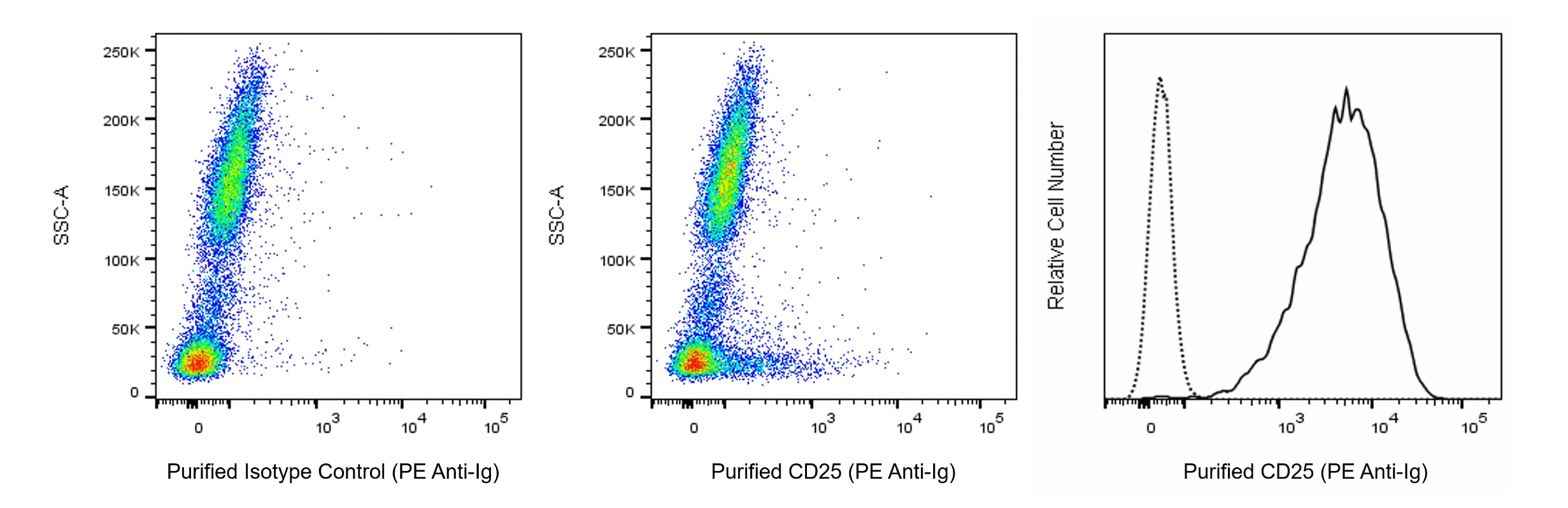
Human (QC Testing)
Mouse BALB/c IgG1, κ
Recombinant Human CD25
Flow cytometry (Routinely Tested)
0.5 mg/ml
V C012
3559
Aqueous buffered solution containing ≤0.09% sodium azide.
RUO


Product Notices

  1. Please refer to www.bdbiosciences.com/us/s/resources for technical protocols.
  2. Since applications vary, each investigator should titrate the reagent to obtain optimal results.
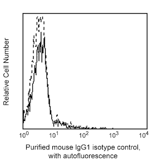
  3. An isotype control should be used at the same concentration as the antibody of interest.
  4. Caution: Sodium azide yields highly toxic hydrazoic acid under acidic conditions. Dilute azide compounds in running water before discarding to avoid accumulation of potentially explosive deposits in plumbing.
  5. Sodium azide is a reversible inhibitor of oxidative metabolism; therefore, antibody preparations containing this preservative agent must not be used in cell cultures nor injected into animals. Sodium azide may be removed by washing stained cells or plate-bound antibody or dialyzing soluble antibody in sodium azide-free buffer. Since endotoxin may also affect the results of functional studies, we recommend the NA/LE (No Azide/Low Endotoxin) antibody format, if available, for in vitro and in vivo use.
  6. Please refer to http://regdocs.bd.com to access safety data sheets (SDS).
568881 Rev. 1
抗体の詳細
Down Arrow Up Arrow
BC96

The BC96 monoclonal antibody specifically binds to human CD25, the low-affinity alpha subunit of the Interleukin-2 Receptor (IL-2Rα), which is also known as TAC antigen. IL2RA encodes CD25 which is a 55 kDa type I transmembrane glycoprotein comprised of an extracellular region with two Complement Control Protein domains (CCP) followed by a transmembrane region and a short cytoplasmic tail. CD25 is constitutively expressed at high levels on natural T regulatory cells and variably expressed on conventional T cells and B cells and their precursors, NK cells, monocytes, and macrophages. CD25 expression can be highly upregulated upon antigenic or mitogenic stimulation of T cells or B cells. A soluble form of CD25 is found in biological fluids due to proteolytic cleavage of the extracellular region of transmembrane CD25. CD25 noncovalently associates with CD122 (IL-2Rβ chain) and CD132 (IL-2Rγ, also known as the common γ chain or γc) to form the high-affinity signal-transducing IL-2R complex (IL-2Rαβγ).  This heterotrimeric receptor mediates biological activities of IL-2 which can act as a cellular activation, growth, and differentiation factor and regulator of cell viability. Analysis of CD25 expression can be used to characterize the nature of normal leucocytes in their resting states or activated during inflammatory or immune responses as well as those present in certain disease states.

568881 Rev. 1
フォーマットの詳細
Down Arrow Up Arrow
Purified
Tissue culture supernatant is purified by either protein A/G or affinity purification methods. Both methods yield antibody in solution that is free of most other soluble proteins, lipids, etc. This format provides pure antibody that is suitable for a number of downstream applications including: secondary labeling for flow cytometry or microscopy, ELISA, Western blot, etc.
Purified
568881 Rev.1
引用&参考文献
Down Arrow Up Arrow

Development References (4)

  1. Chapel A, Bensussan A, Vilmer E, Dormont D. Differential human immunodeficiency virus expression in CD4+ cloned lymphocytes: from viral latency to replication.. J Virol. 1992; 66(6):3966-70. (Immunogen: Flow cytometry). View Reference
  2. Knapp W. W. Knapp .. et al., ed. Leucocyte typing IV : white cell differentiation antigens. Oxford New York: Oxford University Press; 1989:1-1182.
  3. Poszepczynska E, Bagot M, Echchakir H, et al. Functional characterization of an IL-7-dependent CD4(+)CD8alphaalpha(+) Th3-type malignant cell line derived from a patient with a cutaneous T-cell lymphoma.. 2000; 96(3):1056-63. (Clone-specific). View Reference
  4. Schlossman SF. Stuart F. Schlossman .. et al., ed. Leucocyte typing V : white cell differentiation antigens : proceedings of the fifth international workshop and conference held in Boston, USA, 3-7 November, 1993. Oxford: Oxford University Press; 1995.
すべて表示する (4) 表示項目を減らす
568881 Rev. 1

Please refer to Support Documents for Quality Certificates


Global - Refer to manufacturer's instructions for use and related User Manuals and Technical data sheets before using this products as described


Comparisons, where applicable, are made against older BD Technology, manual methods or are general performance claims.  Comparisons are not made against non-BD technologies, unless otherwise noted.

For Research Use Only. Not for use in diagnostic or therapeutic procedures.