-
抗体試薬
- フローサイトメトリー用試薬
-
ウェスタンブロッティング抗体試薬
- イムノアッセイ試薬
-
シングルセル試薬
- BD® AbSeq Assay
- BD Rhapsody™ Accessory Kits
- BD® OMICS-One Immune Profiler Protein Panel
- BD® Single-Cell Multiplexing Kit
- BD Rhapsody™ TCR/BCR Next Multiomic Assays
- BD Rhapsody™ Targeted mRNA Kits
- BD Rhapsody™ Whole Transcriptome Analysis (WTA) Amplification Kit
- BD® OMICS-Guard Sample Preservation Buffer
- BD Rhapsody™ ATAC-Seq Assays
- BD® OMICS-One Protein Panels
-
細胞機能評価のための試薬
-
顕微鏡・イメージング用試薬
-
細胞調製・分離試薬
-
- BD® AbSeq Assay
- BD Rhapsody™ Accessory Kits
- BD® OMICS-One Immune Profiler Protein Panel
- BD® Single-Cell Multiplexing Kit
- BD Rhapsody™ TCR/BCR Next Multiomic Assays
- BD Rhapsody™ Targeted mRNA Kits
- BD Rhapsody™ Whole Transcriptome Analysis (WTA) Amplification Kit
- BD® OMICS-Guard Sample Preservation Buffer
- BD Rhapsody™ ATAC-Seq Assays
- BD® OMICS-One Protein Panels
- Japan (Japanese)
-
Change country/language
Old Browser
Looks like you're visiting us from United States.
Would you like to stay on the current country site or be switched to your country?
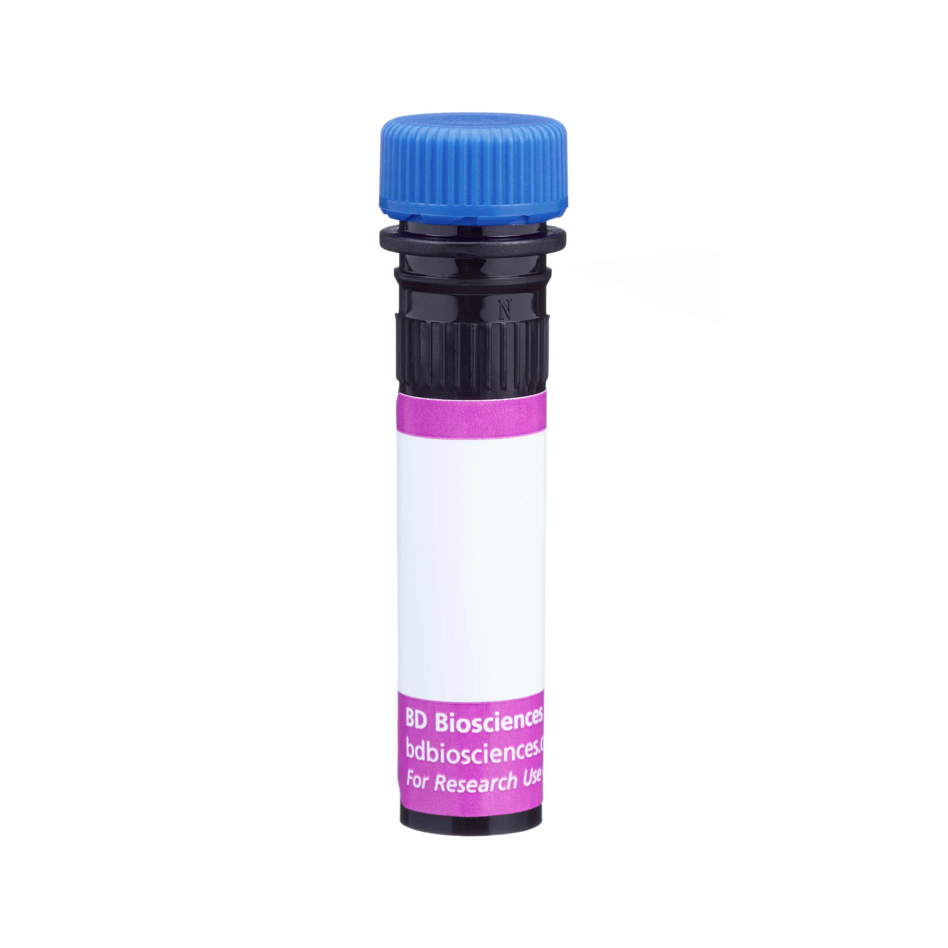
BD Horizon™ BV421 Rat Anti-Mouse TCR Vβ14
クローン 14-2 (RUO)

Flow cytometric analysis of TCR Vβ14 expression on viable Mouse splenic T cells. BALB/c Mouse splenic leukocytes were preincubated with Purified Rat Anti-Mouse CD16/CD32 antibody (Mouse BD Fc Block™) [Cat. No. 553142]. The cells were then stained with PE Hamster Anti-Mouse CD3e antibody (Cat. No. 553064) and with either BD Horizon™ BV421 Mouse IgM, κ Isotype Control (Cat. No. 562708; Left Plot) or BD Horizon™ BV421 Mouse Anti-Mouse TCR Vβ14 antibody (Cat. No. 568788/568789; Right Plot) at 0.25 μg/test. DAPI (4',6-Diamidino-2-Phenylindole, Dihydrochloride) Solution (Cat. No. 564907) was added to cells right before analysis. The bivariate contour plot showing the correlated expression of TCR Vβ14 (or Ig Isotype control staining) versus CD3 was derived from gated events with the forward and side light-scatter characteristics of viable (7-AAD-negative) CD3e-positive splenic lymphocytes. Flow cytometry and data analysis were performed using a BD LSRFortessa™ X-20 Cell Analyzer System and FlowJo™ Software. Data shown on this Technical Data Sheet are not lot specific.

Flow cytometric analysis of TCR Vβ14 expression on viable Mouse splenic T cells. BALB/c Mouse splenic leukocytes were preincubated with Purified Rat Anti-Mouse CD16/CD32 antibody (Mouse BD Fc Block™) [Cat. No. 553142]. The cells were then stained with PE Hamster Anti-Mouse CD3e antibody (Cat. No. 553064) and with either BD Horizon™ BV421 Mouse IgM, κ Isotype Control (Cat. No. 562708; Left Plot) or BD Horizon™ BV421 Mouse Anti-Mouse TCR Vβ14 antibody (Cat. No. 568788/568789; Right Plot) at 0.25 μg/test. DAPI (4',6-Diamidino-2-Phenylindole, Dihydrochloride) Solution (Cat. No. 564907) was added to cells right before analysis. The bivariate contour plot showing the correlated expression of TCR Vβ14 (or Ig Isotype control staining) versus CD3 was derived from gated events with the forward and side light-scatter characteristics of viable (7-AAD-negative) CD3e-positive splenic lymphocytes. Flow cytometry and data analysis were performed using a BD LSRFortessa™ X-20 Cell Analyzer System and FlowJo™ Software. Data shown on this Technical Data Sheet are not lot specific.

Flow cytometric analysis of TCR Vβ14 expression on viable Mouse splenic T cells. BALB/c Mouse splenic leukocytes were preincubated with Purified Rat Anti-Mouse CD16/CD32 antibody (Mouse BD Fc Block™) [Cat. No. 553142]. The cells were then stained with PE Hamster Anti-Mouse CD3e antibody (Cat. No. 553064) and with either BD Horizon™ BV421 Mouse IgM, κ Isotype Control (Cat. No. 562708; Left Plot) or BD Horizon™ BV421 Mouse Anti-Mouse TCR Vβ14 antibody (Cat. No. 568788/568789; Right Plot) at 0.25 μg/test. DAPI (4',6-Diamidino-2-Phenylindole, Dihydrochloride) Solution (Cat. No. 564907) was added to cells right before analysis. The bivariate contour plot showing the correlated expression of TCR Vβ14 (or Ig Isotype control staining) versus CD3 was derived from gated events with the forward and side light-scatter characteristics of viable (7-AAD-negative) CD3e-positive splenic lymphocytes. Flow cytometry and data analysis were performed using a BD LSRFortessa™ X-20 Cell Analyzer System and FlowJo™ Software. Data shown on this Technical Data Sheet are not lot specific.

Flow cytometric analysis of TCR Vβ14 expression on viable Mouse splenic T cells. BALB/c Mouse splenic leukocytes were preincubated with Purified Rat Anti-Mouse CD16/CD32 antibody (Mouse BD Fc Block™) [Cat. No. 553142]. The cells were then stained with PE Hamster Anti-Mouse CD3e antibody (Cat. No. 553064) and with either BD Horizon™ BV421 Mouse IgM, κ Isotype Control (Cat. No. 562708; Left Plot) or BD Horizon™ BV421 Mouse Anti-Mouse TCR Vβ14 antibody (Cat. No. 568788/568789; Right Plot) at 0.25 μg/test. DAPI (4',6-Diamidino-2-Phenylindole, Dihydrochloride) Solution (Cat. No. 564907) was added to cells right before analysis. The bivariate contour plot showing the correlated expression of TCR Vβ14 (or Ig Isotype control staining) versus CD3 was derived from gated events with the forward and side light-scatter characteristics of viable (7-AAD-negative) CD3e-positive splenic lymphocytes. Flow cytometry and data analysis were performed using a BD LSRFortessa™ X-20 Cell Analyzer System and FlowJo™ Software. Data shown on this Technical Data Sheet are not lot specific.


Flow cytometric analysis of TCR Vβ14 expression on viable Mouse splenic T cells. BALB/c Mouse splenic leukocytes were preincubated with Purified Rat Anti-Mouse CD16/CD32 antibody (Mouse BD Fc Block™) [Cat. No. 553142]. The cells were then stained with PE Hamster Anti-Mouse CD3e antibody (Cat. No. 553064) and with either BD Horizon™ BV421 Mouse IgM, κ Isotype Control (Cat. No. 562708; Left Plot) or BD Horizon™ BV421 Mouse Anti-Mouse TCR Vβ14 antibody (Cat. No. 568788/568789; Right Plot) at 0.25 μg/test. DAPI (4',6-Diamidino-2-Phenylindole, Dihydrochloride) Solution (Cat. No. 564907) was added to cells right before analysis. The bivariate contour plot showing the correlated expression of TCR Vβ14 (or Ig Isotype control staining) versus CD3 was derived from gated events with the forward and side light-scatter characteristics of viable (7-AAD-negative) CD3e-positive splenic lymphocytes. Flow cytometry and data analysis were performed using a BD LSRFortessa™ X-20 Cell Analyzer System and FlowJo™ Software. Data shown on this Technical Data Sheet are not lot specific.
Flow cytometric analysis of TCR Vβ14 expression on viable Mouse splenic T cells. BALB/c Mouse splenic leukocytes were preincubated with Purified Rat Anti-Mouse CD16/CD32 antibody (Mouse BD Fc Block™) [Cat. No. 553142]. The cells were then stained with PE Hamster Anti-Mouse CD3e antibody (Cat. No. 553064) and with either BD Horizon™ BV421 Mouse IgM, κ Isotype Control (Cat. No. 562708; Left Plot) or BD Horizon™ BV421 Mouse Anti-Mouse TCR Vβ14 antibody (Cat. No. 568788/568789; Right Plot) at 0.25 μg/test. DAPI (4',6-Diamidino-2-Phenylindole, Dihydrochloride) Solution (Cat. No. 564907) was added to cells right before analysis. The bivariate contour plot showing the correlated expression of TCR Vβ14 (or Ig Isotype control staining) versus CD3 was derived from gated events with the forward and side light-scatter characteristics of viable (7-AAD-negative) CD3e-positive splenic lymphocytes. Flow cytometry and data analysis were performed using a BD LSRFortessa™ X-20 Cell Analyzer System and FlowJo™ Software. Data shown on this Technical Data Sheet are not lot specific.
Flow cytometric analysis of TCR Vβ14 expression on viable Mouse splenic T cells. BALB/c Mouse splenic leukocytes were preincubated with Purified Rat Anti-Mouse CD16/CD32 antibody (Mouse BD Fc Block™) [Cat. No. 553142]. The cells were then stained with PE Hamster Anti-Mouse CD3e antibody (Cat. No. 553064) and with either BD Horizon™ BV421 Mouse IgM, κ Isotype Control (Cat. No. 562708; Left Plot) or BD Horizon™ BV421 Mouse Anti-Mouse TCR Vβ14 antibody (Cat. No. 568788/568789; Right Plot) at 0.25 μg/test. DAPI (4',6-Diamidino-2-Phenylindole, Dihydrochloride) Solution (Cat. No. 564907) was added to cells right before analysis. The bivariate contour plot showing the correlated expression of TCR Vβ14 (or Ig Isotype control staining) versus CD3 was derived from gated events with the forward and side light-scatter characteristics of viable (7-AAD-negative) CD3e-positive splenic lymphocytes. Flow cytometry and data analysis were performed using a BD LSRFortessa™ X-20 Cell Analyzer System and FlowJo™ Software. Data shown on this Technical Data Sheet are not lot specific.
Flow cytometric analysis of TCR Vβ14 expression on viable Mouse splenic T cells. BALB/c Mouse splenic leukocytes were preincubated with Purified Rat Anti-Mouse CD16/CD32 antibody (Mouse BD Fc Block™) [Cat. No. 553142]. The cells were then stained with PE Hamster Anti-Mouse CD3e antibody (Cat. No. 553064) and with either BD Horizon™ BV421 Mouse IgM, κ Isotype Control (Cat. No. 562708; Left Plot) or BD Horizon™ BV421 Mouse Anti-Mouse TCR Vβ14 antibody (Cat. No. 568788/568789; Right Plot) at 0.25 μg/test. DAPI (4',6-Diamidino-2-Phenylindole, Dihydrochloride) Solution (Cat. No. 564907) was added to cells right before analysis. The bivariate contour plot showing the correlated expression of TCR Vβ14 (or Ig Isotype control staining) versus CD3 was derived from gated events with the forward and side light-scatter characteristics of viable (7-AAD-negative) CD3e-positive splenic lymphocytes. Flow cytometry and data analysis were performed using a BD LSRFortessa™ X-20 Cell Analyzer System and FlowJo™ Software. Data shown on this Technical Data Sheet are not lot specific.

Flow cytometric analysis of TCR Vβ14 expression on viable Mouse splenic T cells. BALB/c Mouse splenic leukocytes were preincubated with Purified Rat Anti-Mouse CD16/CD32 antibody (Mouse BD Fc Block™) [Cat. No. 553142]. The cells were then stained with PE Hamster Anti-Mouse CD3e antibody (Cat. No. 553064) and with either BD Horizon™ BV421 Mouse IgM, κ Isotype Control (Cat. No. 562708; Left Plot) or BD Horizon™ BV421 Mouse Anti-Mouse TCR Vβ14 antibody (Cat. No. 568788/568789; Right Plot) at 0.25 μg/test. DAPI (4',6-Diamidino-2-Phenylindole, Dihydrochloride) Solution (Cat. No. 564907) was added to cells right before analysis. The bivariate contour plot showing the correlated expression of TCR Vβ14 (or Ig Isotype control staining) versus CD3 was derived from gated events with the forward and side light-scatter characteristics of viable (7-AAD-negative) CD3e-positive splenic lymphocytes. Flow cytometry and data analysis were performed using a BD LSRFortessa™ X-20 Cell Analyzer System and FlowJo™ Software. Data shown on this Technical Data Sheet are not lot specific.

Flow cytometric analysis of TCR Vβ14 expression on viable Mouse splenic T cells. BALB/c Mouse splenic leukocytes were preincubated with Purified Rat Anti-Mouse CD16/CD32 antibody (Mouse BD Fc Block™) [Cat. No. 553142]. The cells were then stained with PE Hamster Anti-Mouse CD3e antibody (Cat. No. 553064) and with either BD Horizon™ BV421 Mouse IgM, κ Isotype Control (Cat. No. 562708; Left Plot) or BD Horizon™ BV421 Mouse Anti-Mouse TCR Vβ14 antibody (Cat. No. 568788/568789; Right Plot) at 0.25 μg/test. DAPI (4',6-Diamidino-2-Phenylindole, Dihydrochloride) Solution (Cat. No. 564907) was added to cells right before analysis. The bivariate contour plot showing the correlated expression of TCR Vβ14 (or Ig Isotype control staining) versus CD3 was derived from gated events with the forward and side light-scatter characteristics of viable (7-AAD-negative) CD3e-positive splenic lymphocytes. Flow cytometry and data analysis were performed using a BD LSRFortessa™ X-20 Cell Analyzer System and FlowJo™ Software. Data shown on this Technical Data Sheet are not lot specific.

Flow cytometric analysis of TCR Vβ14 expression on viable Mouse splenic T cells. BALB/c Mouse splenic leukocytes were preincubated with Purified Rat Anti-Mouse CD16/CD32 antibody (Mouse BD Fc Block™) [Cat. No. 553142]. The cells were then stained with PE Hamster Anti-Mouse CD3e antibody (Cat. No. 553064) and with either BD Horizon™ BV421 Mouse IgM, κ Isotype Control (Cat. No. 562708; Left Plot) or BD Horizon™ BV421 Mouse Anti-Mouse TCR Vβ14 antibody (Cat. No. 568788/568789; Right Plot) at 0.25 μg/test. DAPI (4',6-Diamidino-2-Phenylindole, Dihydrochloride) Solution (Cat. No. 564907) was added to cells right before analysis. The bivariate contour plot showing the correlated expression of TCR Vβ14 (or Ig Isotype control staining) versus CD3 was derived from gated events with the forward and side light-scatter characteristics of viable (7-AAD-negative) CD3e-positive splenic lymphocytes. Flow cytometry and data analysis were performed using a BD LSRFortessa™ X-20 Cell Analyzer System and FlowJo™ Software. Data shown on this Technical Data Sheet are not lot specific.

Flow cytometric analysis of TCR Vβ14 expression on viable Mouse splenic T cells. BALB/c Mouse splenic leukocytes were preincubated with Purified Rat Anti-Mouse CD16/CD32 antibody (Mouse BD Fc Block™) [Cat. No. 553142]. The cells were then stained with PE Hamster Anti-Mouse CD3e antibody (Cat. No. 553064) and with either BD Horizon™ BV421 Mouse IgM, κ Isotype Control (Cat. No. 562708; Left Plot) or BD Horizon™ BV421 Mouse Anti-Mouse TCR Vβ14 antibody (Cat. No. 568788/568789; Right Plot) at 0.25 μg/test. DAPI (4',6-Diamidino-2-Phenylindole, Dihydrochloride) Solution (Cat. No. 564907) was added to cells right before analysis. The bivariate contour plot showing the correlated expression of TCR Vβ14 (or Ig Isotype control staining) versus CD3 was derived from gated events with the forward and side light-scatter characteristics of viable (7-AAD-negative) CD3e-positive splenic lymphocytes. Flow cytometry and data analysis were performed using a BD LSRFortessa™ X-20 Cell Analyzer System and FlowJo™ Software. Data shown on this Technical Data Sheet are not lot specific.


Regulatory Statusの凡例
Any use of products other than the permitted use without the express written authorization of Becton, Dickinson and Company is strictly prohibited.
Preparation and Storage
推奨アッセイ手順
BD® CompBeads can be used as surrogates to assess fluorescence spillover (compensation). When fluorochrome conjugated antibodies are bound to BD® CompBeads, they have spectral properties very similar to cells. However, for some fluorochromes there can be small differences in spectral emissions compared to cells, resulting in spillover values that differ when compared to biological controls. It is strongly recommended that when using a reagent for the first time, users compare the spillover on cells and BD® CompBeads to ensure that BD® CompBeads are appropriate for your specific cellular application.
For optimal and reproducible results, BD Horizon Brilliant Stain Buffer should be used anytime BD Horizon Brilliant dyes are used in a multicolor flow cytometry panel. Fluorescent dye interactions may cause staining artifacts which may affect data interpretation. The BD Horizon Brilliant Stain Buffer was designed to minimize these interactions. When BD Horizon Brilliant Stain Buffer is used in the multicolor panel, it should also be used in the corresponding compensation controls for all dyes to achieve the most accurate compensation. For the most accurate compensation, compensation controls created with either cells or beads should be exposed to BD Horizon Brilliant Stain Buffer for the same length of time as the corresponding multicolor panel. More information can be found in the Technical Data Sheet of the BD Horizon Brilliant Stain Buffer (Cat. No. 563794/566349) or the BD Horizon Brilliant Stain Buffer Plus (Cat. No. 566385).
Product Notices
- Please refer to www.bdbiosciences.com/us/s/resources for technical protocols.
- Since applications vary, each investigator should titrate the reagent to obtain optimal results.
- An isotype control should be used at the same concentration as the antibody of interest.
- Caution: Sodium azide yields highly toxic hydrazoic acid under acidic conditions. Dilute azide compounds in running water before discarding to avoid accumulation of potentially explosive deposits in plumbing.
- For fluorochrome spectra and suitable instrument settings, please refer to our Multicolor Flow Cytometry web page at www.bdbiosciences.com/colors.
- Please refer to http://regdocs.bd.com to access safety data sheets (SDS).
- For U.S. patents that may apply, see bd.com/patents.
関連製品


The 14-2 antibody reacts with the Vβ 14 T-Cell Receptor (TCR) of mice having the a (e.g., C57BR, C57L, SJL, SWR), b (e.g., A, AKR, BALB/c, CBA, C3H/He, C57BL, C58, DBA/1, DBA/2), and c (e.g., RIII) halpotypes of the Tcrb gene complex. Vβ 14 TCR-expressing T lymphocytes are completely eliminated in mice expressing I-E and the superantigens encoded by Mtv-2 endogenous provirus and/or MMTV-C3H, MMTV-GR, or MMTV-D2.GD exogenous virus. Recognition of these determinants by Vβ 14 TCR-expressing T cells is dependent upon presentation by I-E. Plate bound 14-2 antibody activates Vβ 14 TCR-bearing T cells.
Development References (10)
-
Acha-Orbea H, Shakhov AN, Scarpellino L, et al. Clonal deletion of V beta 14-bearing T cells in mice transgenic for mammary tumour virus. Nature. 1991; 350(6315):207-211. (Biology). View Reference
-
Brady BL, Oropallo MA, Yang-Iott KS, et al. Position-dependent silencing of germline Vß segments on TCRß alleles containing preassembled VßDJßCß1 genes.. J Immunol. 2010; 185(6):3564-73. (Clone-specific: Flow cytometry). View Reference
-
Choi Y, Kappler JW, Marrack P. A superantigen encoded in the open reading frame of the 3' long terminal repeat of mouse mammary tumour virus. Nature. 1991; 350(6315):203-207. (Biology). View Reference
-
Esterházy D, Canesso MCC, Mesin L, et al. Compartmentalized gut lymph node drainage dictates adaptive immune responses.. Nature. 2019; 569(7754):126-130. (Clone-specific: Flow cytometry). View Reference
-
Golovkina TV, Chervonsky A, Dudley JP, Ross SR. Transgenic mouse mammary tumor virus superantigen expression prevents viral infection. Cell. 1992; 69(4):637-645. (Clone-specific). View Reference
-
Hodes RJ, Abe R. Mouse endogenous superantigens: Ms and Mls-like determinants encoded by mouse retroviruses.. Curr Protoc Immunol. 2001; Appendix 1:Appendix 1F. (Biology). View Reference
-
Horowitz JE, Bassing CH. Noncore RAG1 regions promote Vβ rearrangements and αβ T cell development by overcoming inherent inefficiency of Vβ recombination signal sequences.. J Immunol. 2014; 192(4):1609-19. (Clone-specific: Flow cytometry). View Reference
-
Liao NS, Maltzman J, Raulet DH. Positive selection determines T cell receptor V beta 14 gene usage by CD8+ T cells. J Exp Med. 1989; 170(1):135-143. (Immunogen: Flow cytometry, Functional assay, Stimulation). View Reference
-
Xu X, Han L, Zhao G, et al. LRCH1 interferes with DOCK8-Cdc42-induced T cell migration and ameliorates experimental autoimmune encephalomyelitis.. J Exp Med. 2017; 214(1):209-226. (Clone-specific: Flow cytometry). View Reference
-
Zhu ML, Bakhru P, Conley B, et al. Sex bias in CNS autoimmune disease mediated by androgen control of autoimmune regulator.. Nat Commun. 2016; 7:11350. (Clone-specific: Flow cytometry). View Reference
Please refer to Support Documents for Quality Certificates
Global - Refer to manufacturer's instructions for use and related User Manuals and Technical data sheets before using this products as described
Comparisons, where applicable, are made against older BD Technology, manual methods or are general performance claims. Comparisons are not made against non-BD technologies, unless otherwise noted.
For Research Use Only. Not for use in diagnostic or therapeutic procedures.