


製品詳細

BD Pharmingen™
T-cell immunoreceptor with Ig and ITIM domains; VSIG9; VSTM3; WUCAM
Human (QC Testing)
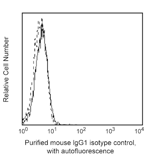
Mouse BALB/c IgG1, κ
Human TIGIT Transfected Cell Line
Blocking, Flow cytometry (Tested During Development)
1.0 mg/ml
AB_3684457
No azide/low endotoxin: Aqueous buffered solution containing no preservative, 0.2µm sterile filtered. Endotoxin level is ≤0.01 EU/µg (≤0.001 ng/µg) of protein as determined by the LAL assay.
RUO
Preparation and Storage
Store undiluted at 4°C. The monoclonal antibody was purified from tissue culture supernatant or ascites by affinity chromatography. This preparation contains no preservatives, thus it should be handled under aseptic conditions.
Product Notices
- Please refer to www.bdbiosciences.com/us/s/resources for technical protocols.
- Since applications vary, each investigator should titrate the reagent to obtain optimal results.
- An isotype control should be used at the same concentration as the antibody of interest.
- Please refer to http://regdocs.bd.com to access safety data sheets (SDS).
関連製品
Stain Buffer (FBS) RUO
サイズ
500 mL
カタログ番号
554656

Stain Buffer (BSA) RUO
サイズ
500 mL
カタログ番号
554657

Purified Mouse IgG1 κ Isotype Control RUO
サイズ
0.1 mg
カタログ番号
556648
PE Streptavidin RUO
サイズ
0.5 mg
カタログ番号
554061
Purified NA/LE Mouse IgG1 κ Isotype Control RUO
サイズ
0.5 mg
カタログ番号
554721

568674 Rev. 1
Please refer to Support Documents for Quality Certificates
Global - Refer to manufacturer's instructions for use and related User Manuals and Technical data sheets before using this products as described
Comparisons, where applicable, are made against older BD Technology, manual methods or are general performance claims. Comparisons are not made against non-BD technologies, unless otherwise noted.
For Research Use Only. Not for use in diagnostic or therapeutic procedures.