Skip to main content Skip to navigation
PE Rat Anti-Mouse CD279 (PD-1)
Flow cytometric analysis of CD279 (PD-1) expression on resting and activated mouse splenocytes. Splenic leucocytes from a C57BL/6 mouse were cultured without (Left Plot) or with (Right Plot) plate-bound Purified NA/LE Hamster Anti-Mouse CD3e antibody (Cat. No. 553057) for 3 days. The resting (Left Plot) or activated (Right Plot) splenocytes were harvested, preincubated with Purified Rat Anti-Mouse CD16/CD32 antibody (Mouse BD Fc Block™) (Cat. No. 553141/553142), and stained with either PE Rat IgG2b, κ Isotype Control (Cat. No. 553989; dashed line histograms) or PE Rat Anti-Mouse CD279 (PD-1) antibody (Cat. No. 566831; solid line histograms) at 1 μg/test. BD Via-Probe™ Cell Viability 7-AAD Solution (Cat. No. 555815/555816) was added to cells right before analysis. The fluorescence histograms showing CD276 (PD-1) expression (or Ig Isotype control staining) were derived from gated events with the forward and side light-scatter characteristics of viable (7-AAD-negative) splenocytes. Flow cytometry and data analysis were performed using a BD LSRFortessa™ X-20 and FlowJo® software. Data shown on this Technical Data Sheet are not lot specific.
製品詳細
Down Arrow Up Arrow


BD Pharmingen™
PD-1; mPD-1; Pdc1; Pdcd1; Ly101
Mouse (QC Testing)
Rat SD, also known as Sprague-Dawley (outbred) IgG2b, κ
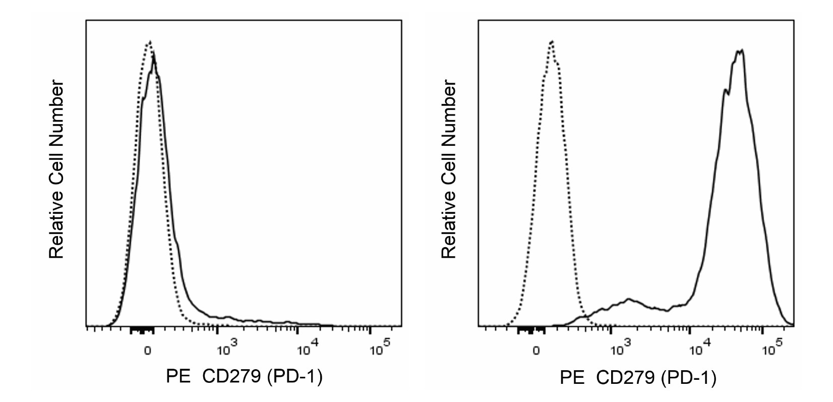
Mouse PD-1 Recombinant Protein
Flow cytometry (Routinely Tested)
0.2 mg/ml
18566
AB_2869890
Aqueous buffered solution containing ≤0.09% sodium azide.
RUO


Preparation and Storage

Store undiluted at 4°C and protected from prolonged exposure to light. Do not freeze. The monoclonal antibody was purified from tissue culture supernatant or ascites by affinity chromatography. The antibody was conjugated with R-PE under optimum conditions, and unconjugated antibody and free PE were removed.

Product Notices

  1. Since applications vary, each investigator should titrate the reagent to obtain optimal results.
  2. An isotype control should be used at the same concentration as the antibody of interest.
  3. Caution: Sodium azide yields highly toxic hydrazoic acid under acidic conditions. Dilute azide compounds in running water before discarding to avoid accumulation of potentially explosive deposits in plumbing.
  4. For fluorochrome spectra and suitable instrument settings, please refer to our Multicolor Flow Cytometry web page at www.bdbiosciences.com/colors.
  5. Please refer to http://regdocs.bd.com to access safety data sheets (SDS).
  6. Please refer to www.bdbiosciences.com/us/s/resources for technical protocols.

関連製品

Purified Rat Anti-Mouse CD16/CD32 (Mouse BD Fc Block™) RUO
サイズ 0.1 mg カタログ番号 553141
sampleImage/
Purified Rat Anti-Mouse CD16/CD32 (Mouse BD Fc Block™) RUO
サイズ 0.5 mg カタログ番号 553142
sampleImage/
Stain Buffer (FBS) RUO
サイズ 500 mL カタログ番号 554656
sampleImage/
Stain Buffer (BSA) RUO
サイズ 500 mL カタログ番号 554657
sampleImage/
PE Rat IgG2b, κ Isotype Control RUO
サイズ 0.1 mg カタログ番号 553989
sampleImage/
Cell Viability Solution RUO
サイズ 500 Tests カタログ番号 555815
sampleImage/
566831 Rev. 1
抗体の詳細
Down Arrow Up Arrow
RMP1-30

The RMP1-30 monoclonal antibody specifically recognizes CD279 which is also known as PD-1 (programmed death-1). CD279 (PD-1) is a ~55 kDa type I transmembrane glycoprotein that is encoded by Pdcd1 which belongs to the CD28/CTLA-4 family within the Ig superfamily. CD279 (PD-1) is comprised of an extracellular region with an IgV-like domain and an intracellular region with an immunoreceptor tyrosine-based inhibitory motif (ITIM) and an immunoreceptor tyrosine-based switch motif (ITSM) that are associated with inhibitory signaling functions. CD279 (PD-1) is transiently expressed on CD4-CD8- thymocytes and developing B lymphocytes at the pro-B-cell stage. It is also expressed on activated myeloid cells, B cells, and T cells including exhausted T cells found in mice during chronic viral infections or cancer. This co-inhibitory receptor reportedly functions in negative regulation of immune responses and thus helps guard against autoimmunity and preserves peripheral tolerance. CD273 (also known as PD-L2 or B7-DC) and CD274 (PD-L1 or B7-H1) are members of the B7 family within the Ig superfamily that serve as ligands for CD279 (PD-1). The RMP1-30 antibody reportedly does not block the binding of CD279 (PD-1) to these ligands.


566831 Rev. 1
フォーマットの詳細
Down Arrow Up Arrow
PE
R-Phycoerythrin (PE), is part of the BD family of Phycobiliprotein dyes. This fluorochrome is a multimeric fluorescent phycobiliprotein with excitation maximum (Ex Max) of 496 nm and 566 nm and an emission maximum (Em Max) at 576 nm. PE is designed to be excited by the Blue (488 nm), Green (532 nm) and Yellow-Green (561 nm) lasers and detected using an optical filter centered near 575 nm (e.g., a 575/26-nm bandpass filter). As PE is excited by multiple lasers, this can result in cross-laser excitation and fluorescence spillover on instruments with various combinations of Blue, Green, and Yellow-Green lasers. Please ensure that your instrument’s configurations (lasers and optical filters) are appropriate for this dye.
altImg
PE
Yellow-Green 488 nm, 532 nm, 561 nm
496 nm, 566 nm
576 nm
566831 Rev.1
引用&参考文献
Down Arrow Up Arrow

Development References (7)

  1. Agata Y, Kawasaki A, Nishimura H, et al. Expression of the PD-1 antigen on the surface of stimulated mouse T and B lymphocytes. Int Immunol. 1996 May; 8(5):765-772. (Biology). View Reference
  2. Gupta PK, Godec J, Wolski D, et al. CD39 Expression Identifies Terminally Exhausted CD8+ T Cells. PLoS Pathogens. 2015; 11(10):e1005177. (Clone-specific: Flow cytometry). View Reference
  3. Kasagi S, Kawano S, Okazaki T, et al. Anti-programmed cell death 1 antibody reduces CD4+PD-1+ T cells and relieves the lupus-like nephritis of NZB/W F1 mice. J Immunol. 2010; 184(5):2337-2347. (Clone-specific: Flow cytometry). View Reference
  4. Liu X, Gibbons RM, Harrington SM, et al. Endogenous tumor-reactive CD8(+) T cells are differentiated effector cells expressing high levels of CD11a and PD-1 but are unable to control tumor growth. Oncoimmunology. 2013; 2(6):e23972. (Clone-specific: Flow cytometry). View Reference
  5. Matsumoto K, Inoue H, Nakano T, et al. B7-DC regulates asthmatic response by an IFN-gamma-dependent mechanism. J Immunol. 2004; 172(4):2530-2541. (Immunogen: Flow cytometry, Functional assay). View Reference
  6. Nishimura H, Agata Y, Kawasaki A, et al. Developmentally regulated expression of the PD-1 protein on the surface of double-negative (CD4-CD8-) thymocytes. Int Immunol. 1996 May; 8(5):773-780. (Biology). View Reference
  7. Tsushima F, Iwai H, Otsuki N, et al. Preferential contribution of B7-H1 to programmed death-1-mediated regulation of hapten-specific allergic inflammatory responses. Eur J Immunol. 2003; 33(10):2773-2782. (Biology). View Reference
すべて表示する (7) 表示項目を減らす
566831 Rev. 1

Please refer to Support Documents for Quality Certificates


Global - Refer to manufacturer's instructions for use and related User Manuals and Technical data sheets before using this products as described


Comparisons, where applicable, are made against older BD Technology, manual methods or are general performance claims.  Comparisons are not made against non-BD technologies, unless otherwise noted.

For Research Use Only. Not for use in diagnostic or therapeutic procedures.