-
抗体試薬
- フローサイトメトリー用試薬
-
ウェスタンブロッティング抗体試薬
- イムノアッセイ試薬
-
シングルセル試薬
- BD® AbSeq Assay
- BD Rhapsody™ Accessory Kits
- BD® OMICS-One Immune Profiler Protein Panel
- BD® Single-Cell Multiplexing Kit
- BD Rhapsody™ TCR/BCR Next Multiomic Assays
- BD Rhapsody™ Targeted mRNA Kits
- BD Rhapsody™ Whole Transcriptome Analysis (WTA) Amplification Kit
- BD® OMICS-Guard Sample Preservation Buffer
- BD Rhapsody™ ATAC-Seq Assays
- BD® OMICS-One Protein Panels
-
細胞機能評価のための試薬
-
顕微鏡・イメージング用試薬
-
細胞調製・分離試薬
-
- BD® AbSeq Assay
- BD Rhapsody™ Accessory Kits
- BD® OMICS-One Immune Profiler Protein Panel
- BD® Single-Cell Multiplexing Kit
- BD Rhapsody™ TCR/BCR Next Multiomic Assays
- BD Rhapsody™ Targeted mRNA Kits
- BD Rhapsody™ Whole Transcriptome Analysis (WTA) Amplification Kit
- BD® OMICS-Guard Sample Preservation Buffer
- BD Rhapsody™ ATAC-Seq Assays
- BD® OMICS-One Protein Panels
- Japan (Japanese)
-
Change country/language
Old Browser
Looks like you're visiting us from United States.
Would you like to stay on the current country site or be switched to your country?
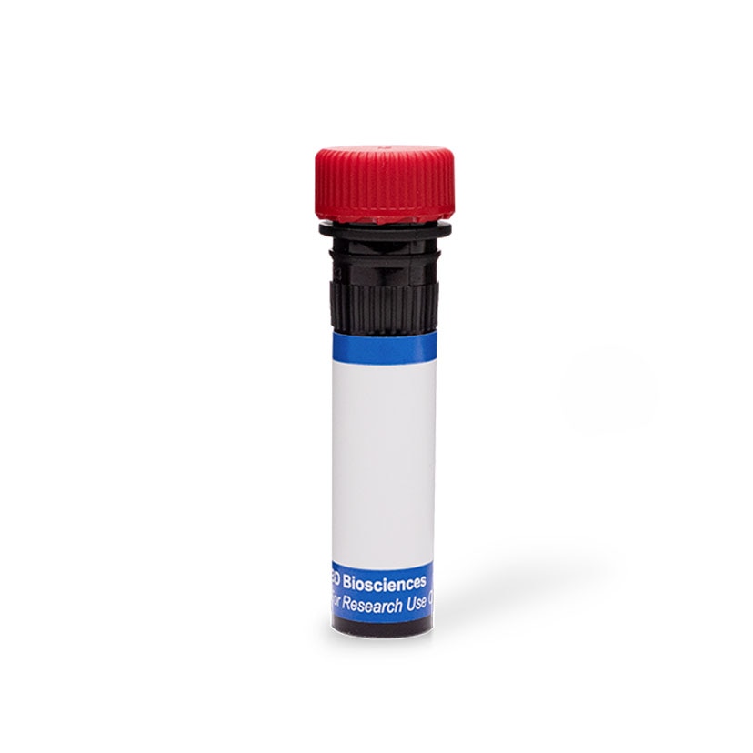
BD Pharmingen™ PE Hamster Anti-Mouse Podoplanin
クローン 8.1.1 (RUO)

Flow cytometric analysis of Podoplanin expressed on mouse C2C12 cells. Cells from the mouse C2C12 (Myoblast, ATCC CRL-1772) cell line were harvested with trypsin-EDTA dissociation solution. The cells were then washed and stained with either PE Hamster IgG2, κ Isotype Control (Cat. No. 550085; dashed line histogram) or PE Hamster Anti-Mouse Podoplanin antibody (Cat. No. 566390; solid line histogram) at 0.25 μg/ml. The histogram showing Podoplanin expression (or Ig Isotype control staining) was derived from gated events with the forward and side light-scatter characteristics of viable C2C12 cells. Flow cytometric analysis was performed using a BD™ Canto II Flow Cytometer System. Data shown on this Technical Data Sheet are not lot specific.


Flow cytometric analysis of Podoplanin expressed on mouse C2C12 cells. Cells from the mouse C2C12 (Myoblast, ATCC CRL-1772) cell line were harvested with trypsin-EDTA dissociation solution. The cells were then washed and stained with either PE Hamster IgG2, κ Isotype Control (Cat. No. 550085; dashed line histogram) or PE Hamster Anti-Mouse Podoplanin antibody (Cat. No. 566390; solid line histogram) at 0.25 μg/ml. The histogram showing Podoplanin expression (or Ig Isotype control staining) was derived from gated events with the forward and side light-scatter characteristics of viable C2C12 cells. Flow cytometric analysis was performed using a BD™ Canto II Flow Cytometer System. Data shown on this Technical Data Sheet are not lot specific.

Flow cytometric analysis of Podoplanin expressed on mouse C2C12 cells. Cells from the mouse C2C12 (Myoblast, ATCC CRL-1772) cell line were harvested with trypsin-EDTA dissociation solution. The cells were then washed and stained with either PE Hamster IgG2, κ Isotype Control (Cat. No. 550085; dashed line histogram) or PE Hamster Anti-Mouse Podoplanin antibody (Cat. No. 566390; solid line histogram) at 0.25 μg/ml. The histogram showing Podoplanin expression (or Ig Isotype control staining) was derived from gated events with the forward and side light-scatter characteristics of viable C2C12 cells. Flow cytometric analysis was performed using a BD™ Canto II Flow Cytometer System. Data shown on this Technical Data Sheet are not lot specific.


BD Pharmingen™ PE Hamster Anti-Mouse Podoplanin

Regulatory Statusの凡例
Any use of products other than the permitted use without the express written authorization of Becton, Dickinson and Company is strictly prohibited.
Preparation and Storage
Product Notices
- Since applications vary, each investigator should titrate the reagent to obtain optimal results.
- An isotype control should be used at the same concentration as the antibody of interest.
- Caution: Sodium azide yields highly toxic hydrazoic acid under acidic conditions. Dilute azide compounds in running water before discarding to avoid accumulation of potentially explosive deposits in plumbing.
- For fluorochrome spectra and suitable instrument settings, please refer to our Multicolor Flow Cytometry web page at www.bdbiosciences.com/colors.
- Although hamster immunoglobulin isotypes have not been well defined, BD Biosciences Pharmingen has grouped Armenian and Syrian hamster IgG monoclonal antibodies according to their reactivity with a panel of mouse anti-hamster IgG mAbs. A table of the hamster IgG groups, Reactivity of Mouse Anti-Hamster Ig mAbs, may be viewed at http://www.bdbiosciences.com/documents/hamster_chart_11x17.pdf.
- Please refer to www.bdbiosciences.com/us/s/resources for technical protocols.
関連製品


最近閲覧済み
The 8.1.1 monoclonal antibody specifically recognizes Podoplanin which is encoded by Pdpn. Podoplanin is a ~43 kDa type I transmembrane glycoprotein that is also known as Glycoprotein 38 (Gp38), OTS-8 (Ots8), Aggrus, RANDAM-2, or T1alpha (T1A). This heavily glycosylated mucin type protein is named for its expression on kidney glomerular epithelial cells known as podocytes. It is also expressed on epithelial and mesothelial cells including intestinal and thymic epithelial cells, alveolar type I cells, fibroblastic reticular cells, lymphatic endothelial cells, macrophages and osteoblasts. Podoplanin plays an essential role in the development of the heart, lymphatic system, and lungs. Podoplanin is involved in actin cytoskeleton organization, and cellular adhesion and migration. It may also play roles in platelet aggregation, promoting inflammatory diseases, tumorigenesis and cancer cell motility and metastasis.

Development References (5)
-
Astarita JL, Acton SE, Turley SJ. Podoplanin: emerging functions in development, the immune system, and cancer.. Front Immunol. 2012; 3:283. (Biology). View Reference
-
Farr A, Nelson A, Hosier S. Characterization of an antigenic determinant preferentially expressed by type I epithelial cells in the murine thymus. J Histochem Cytochem. 1992; 40(5):651-664. (Immunogen: Flow cytometry, Immunofluorescence, Immunohistochemistry, Immunoprecipitation, Western blot). View Reference
-
Farr AG, Berry ML, Kim A, Nelson AJ, Welch MP, Aruffo A. Characterization and cloning of a novel glycoprotein expressed by stromal cells in T-dependent areas of peripheral lymphoid tissues.. J Exp Med. 1992; 176(5):1477-82. (Clone-specific: Electron microscopy, Immunohistochemistry, Western blot). View Reference
-
Malhotra D, Fletcher AL, Astarita J, et al. Transcriptional profiling of stroma from inflamed and resting lymph nodes defines immunological hallmarks.. Nat Immunol. 2012; 13(5):499-510. (Clone-specific: Flow cytometry). View Reference
-
Vigl B, Aebischer D, Nitschké M, et al. Tissue inflammation modulates gene expression of lymphatic endothelial cells and dendritic cell migration in a stimulus-dependent manner.. Blood. 2011; 118(1):205-15. (Clone-specific: Flow cytometry, Fluorescence activated cell sorting). View Reference
Please refer to Support Documents for Quality Certificates
Global - Refer to manufacturer's instructions for use and related User Manuals and Technical data sheets before using this products as described
Comparisons, where applicable, are made against older BD Technology, manual methods or are general performance claims. Comparisons are not made against non-BD technologies, unless otherwise noted.
For Research Use Only. Not for use in diagnostic or therapeutic procedures.
