Skip to main content Skip to navigation
APC-H7 Mouse Anti-Human CD7
Flow cytometric analysis of CD7 expression on human peripheral blood lymphocytes. Whole blood was stained with either APC-H7 Mouse IgG1, κ Isotype Control (Cat. No. 561427; dashed line histogram) or APC-H7 Mouse Anti-Human CD7 antibody (Cat. No. 564020; solid line histogram). The erythrocytes were lysed with BD FACS™ Lysing Solution (Cat. No. 349202). The fluorescence histograms were derived from events with the forward and side light-scatter characteristics of intact lymphocytes. Flow cytometric analysis was performed using a BD™ LSR II Flow Cytometer System.
製品詳細
Down Arrow Up Arrow


BD Pharmingen™
GP40; LEU-9; T-cell leukemia antigen; Tp40; TP41
Human (QC Testing)
Mouse BALB/c IgG1, κ
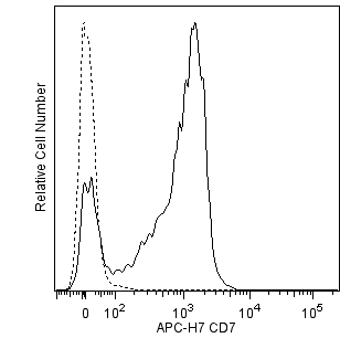
P-CLL and Jurkat Cells
Flow cytometry (Routinely Tested)
5 µl
IV T163
924
AB_2738546
Aqueous buffered solution containing BSA, protein stabilizer, and ≤0.09% sodium azide.
RUO


Preparation and Storage

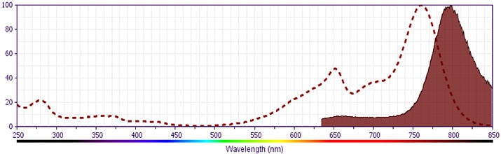
Store undiluted at 4°C and protected from prolonged exposure to light. Do not freeze. The monoclonal antibody was purified from tissue culture supernatant or ascites by affinity chromatography. The antibody was conjugated with APC-H7 under optimum conditions, and unconjugated antibody and APC-H7 were removed.

Product Notices

  1. This reagent has been pre-diluted for use at the recommended Volume per Test. We typically use 1 × 10^6 cells in a 100-µl experimental sample (a test).
  2. An isotype control should be used at the same concentration as the antibody of interest.
  3. Caution: Sodium azide yields highly toxic hydrazoic acid under acidic conditions. Dilute azide compounds in running water before discarding to avoid accumulation of potentially explosive deposits in plumbing.
  4. Source of all serum proteins is from USDA inspected abattoirs located in the United States.
  5. Please observe the following precautions: Absorption of visible light can significantly alter the energy transfer occurring in any tandem fluorochrome conjugate; therefore, we recommend that special precautions be taken (such as wrapping vials, tubes, or racks in aluminum foil) to prevent exposure of conjugated reagents, including cells stained with those reagents, to room illumination.
  6. BD APC-H7 is a tandem conjugate and an analog of APC-Cy7 with the same spectral properties. It has decreased intensity but it is engineered for greater stability and less spillover in the APC channel and consequently offers better performance than APC-Cy7. It has an absorption maximum of approximately 650 nm. When excited by light from a red laser, the APC fluorochrome can transfer energy to the cyanine dye, which then emits at a longer wavelength. The resulting fluorescent emission maximum is approximately 767 nm. BD recommends that a 750-nm longpass filter be used along with a red-sensitive detector such as the Hamamatsu R3896 PMT. As with APC-Cy7 special filters are required when using APC-H7 in conjunction with APC. Note: Although our APC-H7 products demonstrate higher lot-to lot consistency than other APC tandem conjugate products, and every effort is made to minimize the lot-to-lot variation in residual emission from APC, it is strongly recommended that every lot be tested for differences in the amount of compensation required and that individual compensation controls are run for each APC-H7 conjugate.
  7. Although BD APC-H7 is engineered to minimize spillover to the APC channel and is more stable and less affected by light, temperature, and formaldehyde-based fixatives, compared to other APC-cyanine tandem dyes, it is still good practice to minimize as much as possible, any light, temperature and fixative exposure when working with all fluorescent conjugates.
  8. For fluorochrome spectra and suitable instrument settings, please refer to our Multicolor Flow Cytometry web page at www.bdbiosciences.com/colors.
  9. Cy is a trademark of GE Healthcare.
  10. Please refer to www.bdbiosciences.com/us/s/resources for technical protocols.
564020 Rev. 2
抗体の詳細
Down Arrow Up Arrow
M-T701

The M-T701 monoclonal antibody specifically binds to CD7. CD7 is a 40 kDa type I transmembrane glycoprotein that belongs to the immunoglobulin superfamily.  The CD7 antigen is also known as Leu-9, TP41, Tp40, GP40, and T-cell leukemia antigen. It is expressed on thymocytes, T cells, pre-B cells, and NK cells. CD7 is present in reduced density on monocytic cells and cell lines. Functional studies demonstrate that crosslinking of the CD7 can induce transmembrane calcium flux in T cells.


564020 Rev. 2
フォーマットの詳細
Down Arrow Up Arrow
APC-H7
The BD Horizon™ APC-H7 dye is a part of the BD APC red family of dyes. This tandem fluorochrome is comprised of a Allophycocyanin (APC) donor that has excitation maxima (Ex Max) of 659 nm and an acceptor dye, H7, with an emission maximum (Em Max) at 782 nm. APC-H7, driven by BD innovation, is designed to be excited by the Red (627-640 nm) laser and detected using an optical filter centered near 780 nm (e.g., a 760/60 nm bandpass filter). Please ensure that your instrument’s configurations (lasers and optical filters) are appropriate for this dye.
altImg
APC-H7
Red 627-640 nm
659 nm
782 nm
564020 Rev.2
引用&参考文献
Down Arrow Up Arrow

Development References (4)

  1. Pace KE, Lee C, Stewart PL, Baum LG. Restricted receptor segregation into membrane microdomains occurs on human T cells during apoptosis induced by galectin-1. J Immunol. 1999; 163(7):3801-3811. (Clone-specific: Fluorescence microscopy, Functional assay, Immunofluorescence). View Reference
  2. Rabinowich H, Pricop L, Herberman RB, Whiteside TL. Expression and function of CD7 molecule on human natural killer cells. J Immunol. 1994; 152(2):517-526. (Biology). View Reference
  3. Reiter C. Cluster report: CD7. In: Knapp W. W. Knapp .. et al., ed. Leucocyte typing IV : white cell differentiation antigens. Oxford New York: Oxford University Press; 1989:341-342.
  4. Zola H. Leukocyte and stromal cell molecules : the CD markers. Hoboken, N.J.: Wiley-Liss; 2007.
すべて表示する (4) 表示項目を減らす
564020 Rev. 2

Please refer to Support Documents for Quality Certificates


Global - Refer to manufacturer's instructions for use and related User Manuals and Technical data sheets before using this products as described


Comparisons, where applicable, are made against older BD Technology, manual methods or are general performance claims.  Comparisons are not made against non-BD technologies, unless otherwise noted.

For Research Use Only. Not for use in diagnostic or therapeutic procedures.