-
抗体試薬
- フローサイトメトリー用試薬
-
ウェスタンブロッティング抗体試薬
- イムノアッセイ試薬
-
シングルセル試薬
- BD® AbSeq Assay
- BD Rhapsody™ Accessory Kits
- BD® OMICS-One Immune Profiler Protein Panel
- BD® Single-Cell Multiplexing Kit
- BD Rhapsody™ TCR/BCR Next Multiomic Assays
- BD Rhapsody™ Targeted mRNA Kits
- BD Rhapsody™ Whole Transcriptome Analysis (WTA) Amplification Kit
- BD® OMICS-Guard Sample Preservation Buffer
- BD Rhapsody™ ATAC-Seq Assays
- BD® OMICS-One Protein Panels
-
細胞機能評価のための試薬
-
顕微鏡・イメージング用試薬
-
細胞調製・分離試薬
-
- BD® AbSeq Assay
- BD Rhapsody™ Accessory Kits
- BD® OMICS-One Immune Profiler Protein Panel
- BD® Single-Cell Multiplexing Kit
- BD Rhapsody™ TCR/BCR Next Multiomic Assays
- BD Rhapsody™ Targeted mRNA Kits
- BD Rhapsody™ Whole Transcriptome Analysis (WTA) Amplification Kit
- BD® OMICS-Guard Sample Preservation Buffer
- BD Rhapsody™ ATAC-Seq Assays
- BD® OMICS-One Protein Panels
- Japan (Japanese)
-
Change country/language
Old Browser
Looks like you're visiting us from United States.
Would you like to stay on the current country site or be switched to your country?
BD Pharmingen™ Purified Mouse Anti-Human CD324 (E-Cadherin)
クローン 67A4 (RUO)

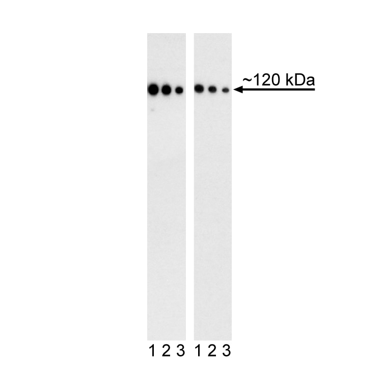
Western blot analysis of CD324 (E-cadherin) expression in human breast adenocarcinoma and human embryonic stem (ES) cells. Cell Lysates from a human breast adenocarcinoma cell line MCF-7 (ATCC HTB-22, left blot) and H9 human ES Cells (WiCell, Madison, WI, right blot) were probed with Purified Mouse Anti-Human CD324 (E-Cadherin) monoclonal antibody at titrations of 0.125 (lanes 1), 0.063 (lanes 2), 0.032 (lanes 3) µg/ml. Proteins were detected using HRP Goat Anti-Mouse Ig (Cat. No. 554002). CD324 (E-cadherin) is identified as a band of ~120 kDa in MCF7 and Human ES cells.

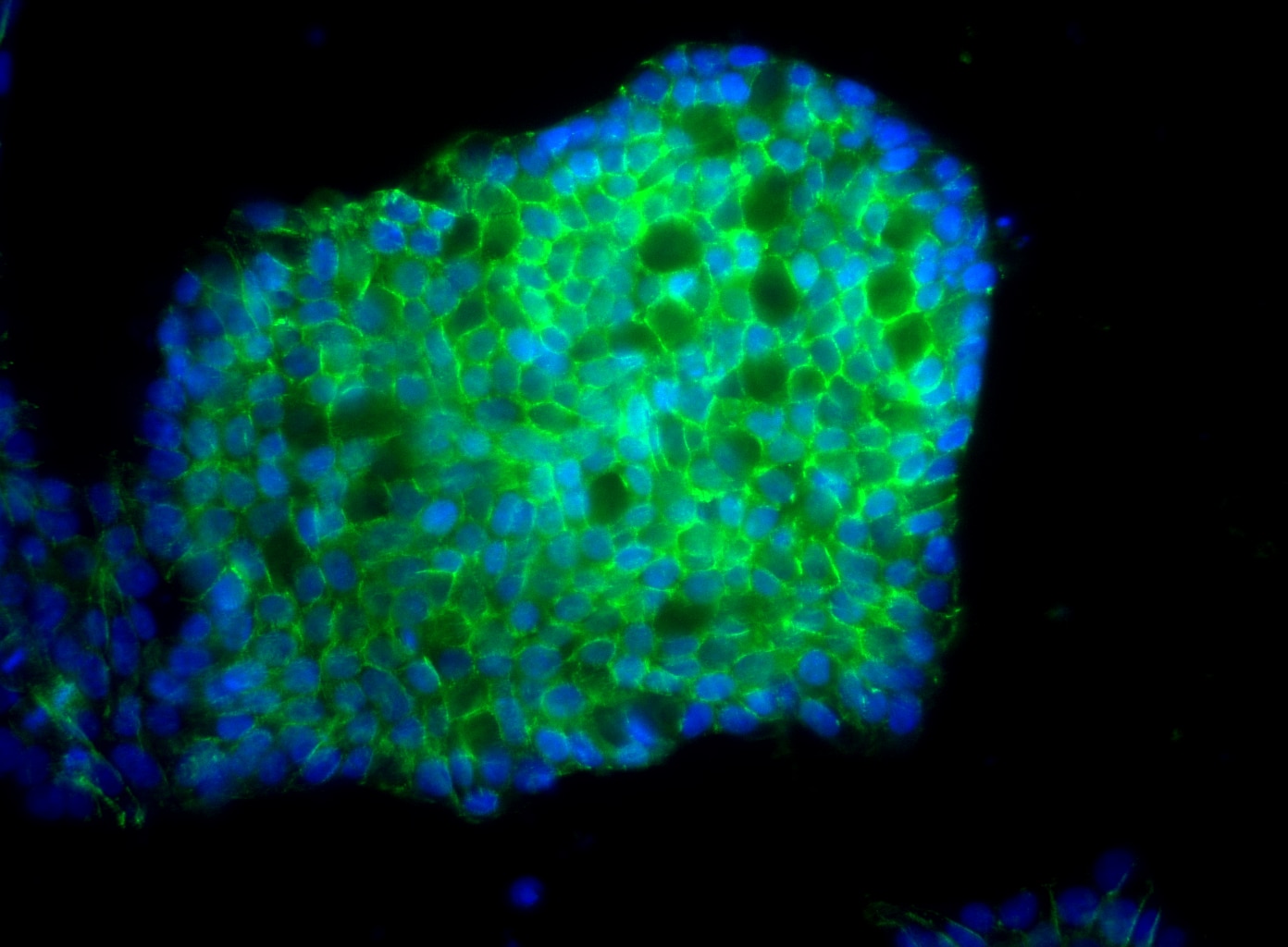
Immunoflourescent staining of CD324 (E-cadherin) on human embryonic stem (ES) cells. H9 human ES cells (WiCell, Madison, WI) passage 45 grown in mTESR™1 media (StemCell Technologies) on BD Matrigel™ hESC-qualified Matrix (Cat. No. 354277) were fixed with BD Cytofix™ Fixation Buffer (Cat. No. 554655). Cells were stained with Purified Mouse Anti-Human CD324 (E-Cadherin) monoclonal antibody (pseudo-colored green) at 2.5 μg/ml. The second-step reagent was Alexa Fluor® 488 goat anti-mouse Ig (Life Technologies), and cell nuclei were stained with DAPI (pseudo-colored blue). The images were captured on a BD Pathway™ 435 Cell Analyzer and merged using BD Attovision™ software.


Western blot analysis of CD324 (E-cadherin) expression in human breast adenocarcinoma and human embryonic stem (ES) cells. Cell Lysates from a human breast adenocarcinoma cell line MCF-7 (ATCC HTB-22, left blot) and H9 human ES Cells (WiCell, Madison, WI, right blot) were probed with Purified Mouse Anti-Human CD324 (E-Cadherin) monoclonal antibody at titrations of 0.125 (lanes 1), 0.063 (lanes 2), 0.032 (lanes 3) µg/ml. Proteins were detected using HRP Goat Anti-Mouse Ig (Cat. No. 554002). CD324 (E-cadherin) is identified as a band of ~120 kDa in MCF7 and Human ES cells.
Immunoflourescent staining of CD324 (E-cadherin) on human embryonic stem (ES) cells. H9 human ES cells (WiCell, Madison, WI) passage 45 grown in mTESR™1 media (StemCell Technologies) on BD Matrigel™ hESC-qualified Matrix (Cat. No. 354277) were fixed with BD Cytofix™ Fixation Buffer (Cat. No. 554655). Cells were stained with Purified Mouse Anti-Human CD324 (E-Cadherin) monoclonal antibody (pseudo-colored green) at 2.5 μg/ml. The second-step reagent was Alexa Fluor® 488 goat anti-mouse Ig (Life Technologies), and cell nuclei were stained with DAPI (pseudo-colored blue). The images were captured on a BD Pathway™ 435 Cell Analyzer and merged using BD Attovision™ software.

Western blot analysis of CD324 (E-cadherin) expression in human breast adenocarcinoma and human embryonic stem (ES) cells. Cell Lysates from a human breast adenocarcinoma cell line MCF-7 (ATCC HTB-22, left blot) and H9 human ES Cells (WiCell, Madison, WI, right blot) were probed with Purified Mouse Anti-Human CD324 (E-Cadherin) monoclonal antibody at titrations of 0.125 (lanes 1), 0.063 (lanes 2), 0.032 (lanes 3) µg/ml. Proteins were detected using HRP Goat Anti-Mouse Ig (Cat. No. 554002). CD324 (E-cadherin) is identified as a band of ~120 kDa in MCF7 and Human ES cells.

Immunoflourescent staining of CD324 (E-cadherin) on human embryonic stem (ES) cells. H9 human ES cells (WiCell, Madison, WI) passage 45 grown in mTESR™1 media (StemCell Technologies) on BD Matrigel™ hESC-qualified Matrix (Cat. No. 354277) were fixed with BD Cytofix™ Fixation Buffer (Cat. No. 554655). Cells were stained with Purified Mouse Anti-Human CD324 (E-Cadherin) monoclonal antibody (pseudo-colored green) at 2.5 μg/ml. The second-step reagent was Alexa Fluor® 488 goat anti-mouse Ig (Life Technologies), and cell nuclei were stained with DAPI (pseudo-colored blue). The images were captured on a BD Pathway™ 435 Cell Analyzer and merged using BD Attovision™ software.


BD Pharmingen™ Purified Mouse Anti-Human CD324 (E-Cadherin)

BD Pharmingen™ Purified Mouse Anti-Human CD324 (E-Cadherin)

Regulatory Statusの凡例
Any use of products other than the permitted use without the express written authorization of Becton, Dickinson and Company is strictly prohibited.
Preparation and Storage
Product Notices
- Since applications vary, each investigator should titrate the reagent to obtain optimal results.
- Please refer to www.bdbiosciences.com/us/s/resources for technical protocols.
- Caution: Sodium azide yields highly toxic hydrazoic acid under acidic conditions. Dilute azide compounds in running water before discarding to avoid accumulation of potentially explosive deposits in plumbing.
- Sodium azide is a reversible inhibitor of oxidative metabolism; therefore, antibody preparations containing this preservative agent must not be used in cell cultures nor injected into animals. Sodium azide may be removed by washing stained cells or plate-bound antibody or dialyzing soluble antibody in sodium azide-free buffer. Since endotoxin may also affect the results of functional studies, we recommend the NA/LE (No Azide/Low Endotoxin) antibody format, if available, for in vitro and in vivo use.
- Alexa Fluor® is a registered trademark of Molecular Probes, Inc., Eugene, OR.
- All other brands are trademarks of their respective owners.
関連製品


The 67A4 monoclonal antibody specifically recognizes the extracellular domain of human E-Cadherin (CD324). E-Cadherin is a 120-kDa transmembrane glycoprotein that is localized in the adherens junctions of epithelial cells. There it interacts with the cytoskeleton through the associated cytoplasmic catenin proteins. In addition to being a calcium-dependent adhesion molecule, E-Cadherin is also a critical regulator of epithelial junction formation. Its association with catenins is necessary for cell-to-cell adhesion. These E-Cadherin/catenin complexes associate with cortical actin bundles at both the zonula adherens and the lateral adhesion plaques. Tyrosine phosphorylation can disrupt these complexes, leading to changes in cell adhesion properties. E-Cadherin expression is often down-regulated in highly invasive, poorly differentiated carcinomas. Increased expression of E-Cadherin in these cells reduces their invasiveness. Thus, loss of expression or function of E-Cadherin appears to be an important step in tumorigenic progression. Pluripotent stem cells express E-Cadherin. Upon differentiation, an epithelial to mesenchymal transition results in the loss of E-cadherin expression and a gain in the expression of N-cadherin.
Development References (5)
-
Behrens J, Vakaet L, Friis R, et al. Loss of epithelial differentiation and gain of invasiveness correlates with tyrosine phosphorylation of the E-cadherin/beta-catenin complex in cells transformed with a temperature-sensitive v-SRC gene.. J Cell Biol. 1993; 120(3):757-66. (Biology). View Reference
-
Bühring HJ, Müller T, Herbst R, et al. The adhesion molecule E-cadherin and a surface antigen recognized by the antibody 9C4 are selectively expressed on erythroid cells of defined maturational stages.. Leukemia. 1996; 10(1):106-16. (Clone-specific: Flow cytometry). View Reference
-
Cepek KL, Shaw SK, Parker CM, et al. Adhesion between epithelial cells and T lymphocytes mediated by E-cadherin and the alpha E beta 7 integrin.. Nature. 1994; 372(6502):190-3. (Biology). View Reference
-
D'Amour KA, Agulnick AD, Eliazer S, Kelly OG, Kroon E, Baetge EE. Efficient differentiation of human embryonic stem cells to definitive endoderm.. Nat Biotechnol. 2005; 23(12):1534-41. (Biology). View Reference
-
Takeichi M. The cadherins: cell-cell adhesion molecules controlling animal morphogenesis.. Development. 1988; 102(4):639-55. (Biology). View Reference
Please refer to Support Documents for Quality Certificates
Global - Refer to manufacturer's instructions for use and related User Manuals and Technical data sheets before using this products as described
Comparisons, where applicable, are made against older BD Technology, manual methods or are general performance claims. Comparisons are not made against non-BD technologies, unless otherwise noted.
For Research Use Only. Not for use in diagnostic or therapeutic procedures.