Skip to main content Skip to navigation
PE Rat anti-Mouse CD25
Flow cytometric analysis of PE-conjugated anti-mouse CD25 on mouse splenocytes. Concanavlin A activated murine splenocytes were  stained with either PE anti-CD25(clone 7D4, Cat. No. 558642, solid line) or a PE rat IgM isotype control (catalog number 553943, dashed line) and analyzed by flow cytometry.  Flow cytometry was performed on a BD FACSCalibur™ System and the histograms were derived from the gated events based on light scattering characteristics of viable splenocytes.
製品詳細
Down Arrow Up Arrow


BD Pharmingen™
IL-2 Receptor α chain, p55
Mouse (QC Testing)
Rat LEW, also known as Lewis IgM, κ
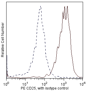
IL-2-dependent BALB/c mouse helper T-cell clone HT-2
Flow cytometry (Routinely Tested)
0.2 mg/ml
AB_1645250
Aqueous buffered solution containing ≤0.09% sodium azide.
RUO


Preparation and Storage

Store undiluted at 4°C and protected from prolonged exposure to light. Do not freeze. The antibody was conjugated with R-PE under optimum conditions, and unconjugated antibody and free PE were removed.

Product Notices

  1. Since applications vary, each investigator should titrate the reagent to obtain optimal results.
  2. Caution: Sodium azide yields highly toxic hydrazoic acid under acidic conditions. Dilute azide compounds in running water before discarding to avoid accumulation of potentially explosive deposits in plumbing.
  3. For fluorochrome spectra and suitable instrument settings, please refer to our Multicolor Flow Cytometry web page at www.bdbiosciences.com/colors.
  4. Please refer to www.bdbiosciences.com/us/s/resources for technical protocols.
558642 Rev. 1
抗体の詳細
Down Arrow Up Arrow
7D4

The 7D4 monoclonal antibody specifically binds to CD25, the low affinity IL-2 Receptor (IL-2Rα, p55) expressed on activated T and B lymphocytes from all mouse strains tested. IL-2Rα by itself is not a signaling receptor. However, it can combine with IL-2 Receptor β (CD122) and γ (CD132) chains to form high-affinity, signaling receptor complexes for IL-2. Resting T and B lymphocytes as well as resting and activated NK cells do not express IL-2Rα.  CD25 is transiently expressed at a low level during normal B-cell development in the bone marrow on the CD45R/B220low TdT- sIg- Pre-B/Pre-B-II and CD45R/B220low TdT- sIgM+ sIgD- immature B stages, but not on the CD45R/B220low TdT+ sIg- Pro-B/Pre-B-I stage nor on CD45R/B220high TdTsIgM+ sIgD+ mature B cells.  It is expressed at a higher level during a very early stage of T-cell development in fetal and adult thymus. Peripheral CD25+ CD4+ T lymphocytes, called regulatory T (Treg) cells, are involved in the maintenance of self-tolerance. It has also been reported that dendritic cells express CD25, which is recognized by mAb 7D4. The 3C7 antibody recognizes an epitope of CD25 which is distinct from those recognized by mAbs 7D4 and PC61, and it blocks binding of IL-2 to CD25.

558642 Rev. 1
フォーマットの詳細
Down Arrow Up Arrow
PE
R-Phycoerythrin (PE), is part of the BD family of Phycobiliprotein dyes. This fluorochrome is a multimeric fluorescent phycobiliprotein with excitation maximum (Ex Max) of 496 nm and 566 nm and an emission maximum (Em Max) at 576 nm. PE is designed to be excited by the Blue (488 nm), Green (532 nm) and Yellow-Green (561 nm) lasers and detected using an optical filter centered near 575 nm (e.g., a 575/26-nm bandpass filter). As PE is excited by multiple lasers, this can result in cross-laser excitation and fluorescence spillover on instruments with various combinations of Blue, Green, and Yellow-Green lasers. Please ensure that your instrument’s configurations (lasers and optical filters) are appropriate for this dye.
altImg
PE
Yellow-Green 488 nm, 532 nm, 561 nm
496 nm, 566 nm
576 nm
558642 Rev.1
引用&参考文献
Down Arrow Up Arrow

Development References (15)

  1. Chen J, Ma A, Young F, Alt FW. IL-2 receptor alpha chain expression during early B lymphocyte differentiation. Int Immunol. 1994; 6(8):1265-1268. (Biology). View Reference
  2. Crowley M, Inaba K, Witmer-Pack M, Steinman RM. The cell surface of mouse dendritic cells: FACS analyses of dendritic cells from different tissues including thymus. Cell Immunol. 1989; 118(1):108-125. (Biology). View Reference
  3. Garni-Wagner BA, Witte PL, Tutt MM, et al. Natural killer cells in the thymus. Studies in mice with severe combined immune deficiency. J Immunol. 1990; 144(3):796-803. (Biology). View Reference
  4. Godfrey DI, Kennedy J, Mombaerts P, Tonegawa S, Zlotnik A. Onset of TCR-β gene rearrangement and role of TCR-β expression during CD3-CD4-CD8- thymocyte differentiation. J Immunol. 1994; 152(10):4783-4792. (Biology). View Reference
  5. Habu S, Okumura K, Diamantstein T, Shevach EM. Expression of interleukin 2 receptor on murine fetal thymocytes. Eur J Immunol. 1985; 15(5):456-460. (Biology). View Reference
  6. Lorenzo F, Jaulin C, Vita N, et al. Structure-function study of the p55 subunit of murine IL-2 receptor by epitope mapping. J Immunol. 1991; 147(9):2970-2977. (Biology). View Reference
  7. Malek TR, Robb RJ, Shevach EM. Identification and initial characterization of a rat monoclonal antibody reactive with the murine interleukin 2 receptor-ligand complex. Proc Natl Acad Sci U S A. 1983; 80(18):5694-5698. (Immunogen). View Reference
  8. Malek TR, Schmidt JA, Shevach EM. The murine IL 2 receptor. III. Cellular requirements for the induction of IL 2 receptor expression on T cell subpopulations. J Immunol. 1985; 134(4):2405-2413. (Biology). View Reference
  9. Moreau JL, Nabholz M, Diamantstein T, Malek T, Shevach E, Theze J. Monoclonal antibodies identify three epitope clusters on the mouse p55 subunit of the interleukin 2 receptor: relationship to the interleukin 2-binding site. Eur J Immunol. 1987; 17(7):929-935. (Biology). View Reference
  10. Ortega G, Robb RJ, Shevach EM, Malek TR. The murine IL 2 receptor. I. Monoclonal antibodies that define distinct functional epitopes on activated T cells and react with activated B cells. J Immunol. 1984; 133(4):1970-1975. (Clone-specific). View Reference
  11. Pollard AM, Lipscomb MF. Characterization of murine lung dendritic cells: similarities to Langerhans cells and thymic dendritic cells. J Exp Med. 1990; 172(1):159-167. (Biology). View Reference
  12. Read S, Malmstrom V, Powrie F. Cytotoxic T lymphocyte-associated antigen 4 plays an essential role in the function of CD25(+)CD4(+) regulatory cells that control intestinal inflammation. J Exp Med. 2000; 192(2):295-302. (Biology). View Reference
  13. Rolink A, Grawunder U, Winkler TH, Karasuyama H, Melchers F. IL-2 receptor alpha chain (CD25, TAC) expression defines a crucial stage in pre-B cell development. Int Immunol. 1994; 6(8):1257-1264. (Biology). View Reference
  14. Takahashi T, Tagami T, Yamazaki S, et al. Immunologic self-tolerance maintained by CD25(+)CD4(+) regulatory T cells constitutively expressing cytotoxic T lymphocyte-associated antigen 4. J Exp Med. 2000; 192(2):303-309. (Biology). View Reference
  15. Taniguchi T, Minami Y. The IL-2/IL-2 receptor system: a current overview. Cell. 1993; 73(1):5-8. (Biology). View Reference
すべて表示する (15) 表示項目を減らす
558642 Rev. 1

Please refer to Support Documents for Quality Certificates


Global - Refer to manufacturer's instructions for use and related User Manuals and Technical data sheets before using this products as described


Comparisons, where applicable, are made against older BD Technology, manual methods or are general performance claims.  Comparisons are not made against non-BD technologies, unless otherwise noted.

For Research Use Only. Not for use in diagnostic or therapeutic procedures.