Skip to main content Skip to navigation
Purified NA/LE Hamster Anti-Mouse MCP-1
製品詳細
Down Arrow Up Arrow


BD Pharmingen™
Mouse (QC Testing)
Armenian Hamster IgG1, κ
Heparin-purified CHO-expressed mouse MCP-1
ELISA (Routinely Tested), Intracellular staining (flow cytometry), Neutralization (Tested During Development)
1.0 mg/ml
AB_395392
No azide/low endotoxin: Aqueous buffered solution containing no preservative, 0.2µm sterile filtered. Endotoxin level is ≤0.01 EU/µg (≤0.001 ng/µg) of protein as determined by the LAL assay.
RUO


Preparation and Storage

Store undiluted at 4°C. The monoclonal antibody was purified from tissue culture supernatant or ascites by affinity chromatography. This preparation contains no preservatives, thus it should be handled under aseptic conditions.

推奨アッセイ手順

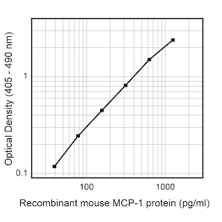
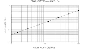
ELISA:  The purified format of the 2H5 antibody (Cat. No. 551217) is useful as a capture antibody in a sandwich ELISA for measuring mouse MCP-1 protein levels when paired with biotin conjugated clone 4E2/MCP (Cat. No, 554444) as a detection reagent and recombinant mouse MCP-1 (Cat. No. 554590) as the standard. For testing mouse MCP-1 in complex biological fluids, such as serum or plasma, investigators are encouraged to consider using the BD OptEIA™ Mouse MCP-1 Set (Cat. No. 555260).

Neutralization: Investigators are advised that this material is not routinely tested for the Neutralization application and are highly encouraged to both titrate this material and include appropriate controls in relevant experiments. This antibody has previously been reported to be useful for the neutralization of recombinant mouse MCP-1 when measured in a calcium flux assay using 100-200 ng/mL recombinant mouse MCP-1 (Cat. No. 554590) to stimulate THP-1 indicator cells with the following representative ranges:

50% Neutralization (ND50) at 2 - 30 μg/mL

90% Neutralization at 40 - 300 μg/mL

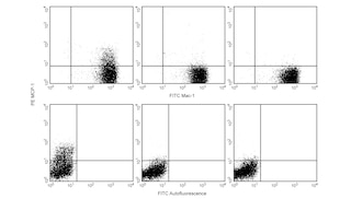
Flow Cytometry:  The 2H5 antibody can be useful for immunofluorescent staining and flow cytometric analysis to identify and enumerate MCP-1 producing cells within mixed cell populations. The PE-conjugated 2H5 antibody (Cat. No. 554443) is especially suitable for these experiments.

Product Notices

  1. Since applications vary, each investigator should titrate the reagent to obtain optimal results.
  2. Please refer to www.bdbiosciences.com/us/s/resources for technical protocols.

関連製品

Purified NA/LE Hamster IgG1 κ Isotype Control RUO
サイズ 0.5 mg カタログ番号 553968
sampleImage/
PE Hamster Anti-Mouse/Rat MCP-1 RUO
サイズ 0.1 mg カタログ番号 554443
sampleImage/
Biotin Hamster Anti-Mouse MCP-1 RUO
サイズ 0.5 mg カタログ番号 554444
sampleImage/
Recombinant Mouse MCP-1 RUO
サイズ 5 µg カタログ番号 554590
sampleImage/
Purified Hamster Anti-Mouse MCP-1 RUO
サイズ 1 mg カタログ番号 551217
sampleImage/
Mouse MCP-1 ELISA Set RUO
サイズ 20 Plate(s) カタログ番号 555260
sampleImage/
554440 Rev. 2
抗体の詳細
Down Arrow Up Arrow
2H5

The 2H5 antibody reacts with mouse and rat monocyte chemoattractant protein (MCP-1), formerly termed JE. This antibody also recognizes human MCP-1, but shows no reactivity with the closely related mouse β chemokines, TCA3 and MIP-1β. The immunogen used to generate the 2H5 hybridoma was heparin-purified CHO-expressed mouse MCP-1. This is a neutralizing antibody.

554440 Rev. 2
フォーマットの詳細
Down Arrow Up Arrow
NA/LE
NA/LE refers to the culture and purification methods and buffer used to produce purified antibodies with no azide and low endotoxin: Aqueous buffered solution containing no preservative, 0.2µm sterile filtered. Endotoxin level is ≤0.01 EU/µg (≤0.001 ng/µg) of protein as determined by the LAL assay.NA/LE are perfectly suited to be used in culture or in vivo (for nonhuman studies) for functional assays — blocking, neutralizing, activation or depletion — where the presence of azide may damage cells or exogenous endotoxin may signal or activate cells.
NA/LE
554440 Rev.2
引用&参考文献
Down Arrow Up Arrow

Development References (3)

  1. Boring L, Gosling J, Monteclaro FS, Lusis AJ, Tsou CL, Charo IF. Molecular cloning and functional expression of murine JE (monocyte chemoattractant protein 1) and murine macrophage inflammatory protein 1alpha receptors: evidence for two closely linked C-C chemokine receptors on chromosome 9. J Biol Chem. 1996; 271(13):7551-7558. (Clone-specific). View Reference
  2. Luo Y, Laning J, Hayashi M, Hancock PR, Rollins B, Dorf ME. Serologic analysis of the mouse beta chemokine JE/monocyte chemoattractant protein-1. J Immunol. 1994; 153(8):3708-3716. (Clone-specific). View Reference
  3. Prussin C, Metcalfe DD. Detection of intracytoplasmic cytokine using flow cytometry and directly conjugated anti-cytokine antibodies. J Immunol Methods. 1995; 188(1):117-128. (Methodology: Apoptosis). View Reference
554440 Rev. 2

Please refer to Support Documents for Quality Certificates


Global - Refer to manufacturer's instructions for use and related User Manuals and Technical data sheets before using this products as described


Comparisons, where applicable, are made against older BD Technology, manual methods or are general performance claims.  Comparisons are not made against non-BD technologies, unless otherwise noted.

For Research Use Only. Not for use in diagnostic or therapeutic procedures.