Skip to main content Skip to navigation
Biotin Hamster Anti-Mouse γδ T-Cell Receptor
Two-color analysis of the expression of γδ TCR on peripheral T lymphocytes. C57BL/6 lymph node cells were incubated simultaneously with Biotin Hamster Anti-Mouse γδ T-Cell Receptor (Cat. No. 553176, right panel) and 
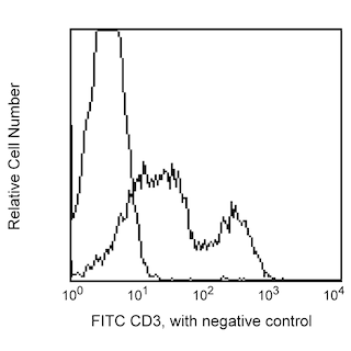
FITC Rat Anti-Mouse CD3 Molecular Complex (Cat. No. 555274), followed by PE Streptavidin (Cat. No. 554061). Fluorescence dot plots were derived from gated events based on the forward and side light-scattering of viable lymphocytes. Flow cytometry was performed on a BD FACScan™.
製品詳細
Down Arrow Up Arrow


BD Pharmingen™
Tcrd; T-cell receptor delta chain; Tcr delta
Mouse (QC Testing)
Armenian Hamster IgG2, κ
C57BL/6 Mouse Intestinal Intraepithelial Lymphocytes
Flow cytometry (Routinely Tested), Immunohistochemistry-frozen (Reported)
0.5 mg/ml
AB_394687
Aqueous buffered solution containing ≤0.09% sodium azide.
RUO


Preparation and Storage

Store undiluted at 4°C. The monoclonal antibody was purified from tissue culture supernatant or ascites by affinity chromatography. The antibody was conjugated with biotin under optimum conditions, and unreacted biotin was removed.

推奨アッセイ手順

For flow cytometry of cell suspensions from peripheral lymphoid tissues, it is recommended that multicolor staining be performed to distinguish T lymphocytes from non-T cells.

Product Notices

  1. Since applications vary, each investigator should titrate the reagent to obtain optimal results.
  2. An isotype control should be used at the same concentration as the antibody of interest.
  3. Caution: Sodium azide yields highly toxic hydrazoic acid under acidic conditions. Dilute azide compounds in running water before discarding to avoid accumulation of potentially explosive deposits in plumbing.
  4. Although hamster immunoglobulin isotypes have not been well defined, BD Biosciences Pharmingen has grouped Armenian and Syrian hamster IgG monoclonal antibodies according to their reactivity with a panel of mouse anti-hamster IgG mAbs. A table of the hamster IgG groups, Reactivity of Mouse Anti-Hamster Ig mAbs, may be viewed at http://www.bdbiosciences.com/documents/hamster_chart_11x17.pdf.
  5. For fluorochrome spectra and suitable instrument settings, please refer to our Multicolor Flow Cytometry web page at www.bdbiosciences.com/colors.
  6. Please refer to www.bdbiosciences.com/us/s/resources for technical protocols.
553176 Rev. 16
抗体の詳細
Down Arrow Up Arrow
GL3

The GL3 monoclonal antibody specifically binds to a common epitope of the δ chain of the T-cell Receptor (TCR) complex on γδ TCR-expressing T lymphocytes and NK-T cells of all mouse strains tested. It does not react with αβ TCR-bearing T cells. In the mouse, cells expressing the γδ TCR are found in the thymus, intestinal epithelium, epidermis, dermis, pulmonsry epithelium, peritoneum, liver, and peripheral lymphoid organs.

553176 Rev. 16
フォーマットの詳細
Down Arrow Up Arrow
Biotin
Biotin is a ubiquitous co-factor (also known as Vitamin B7) that has many properties that make it extremely useful for molecular biology. Biotin has an extremely high affinity for the Avidin family of proteins (Kd = 10-15 M), making it the perfect tool to link two molecules. Biotin labeled antibodies can be combined with any number of Avidin-conjugated probes in order to customize an assay to a particular need. This is especially useful in the case of magnetic cell separation using streptavidin/magnetic bead conjugates, or in the case of flow cytometry using streptavidin/fluorophore conjugates.
Biotin
553176 Rev.16
引用&参考文献
Down Arrow Up Arrow

Development References (12)

  1. Goodman T, LeCorre R, Lefrancois L. A T-cell receptor gamma delta-specific monoclonal antibody detects a V gamma 5 region polymorphism. Immunogenetics. 1992; 35(1):65-68. (Clone-specific: Flow cytometry). View Reference
  2. Goodman T, Lefrancois L. Intraepithelial lymphocytes. Anatomical site, not T cell receptor form, dictates phenotype and function. J Exp Med. 1989; 170(5):1569-1581. (Clone-specific: Flow cytometry). View Reference
  3. Kaufmann SH, Blum C, Yamamoto S. Crosstalk between alpha/beta T cells and gamma/delta T cells in vivo: activation of alpha/beta T-cell responses after gamma/delta T-cell modulation with the monoclonal antibody GL3. Proc Natl Acad Sci U S A. 1993; 90(20):9620-9624. (Clone-specific: Flow cytometry). View Reference
  4. King DP, Hyde DM, Jackson KA, et al. Cutting edge: protective response to pulmonary injury requires gamma delta T lymphocytes. J Immunol. 1999; 162(9):5033-5036. (Biology: Flow cytometry). View Reference
  5. Lefrancois L. Phenotypic complexity of intraepithelial lymphocytes of the small intestine. J Immunol. 1991; 147(6):1746-1751. (Biology: Flow cytometry). View Reference
  6. MacDonald HR, Schreyer M, Howe RC, Bron C. Selective expression of CD8 alpha (Ly-2) subunit on activated thymic gamma/delta cells. Eur J Immunol. 1990; 20(4):927-930. (Biology: Flow cytometry). View Reference
  7. Mosley RL, Klein JR. Peripheral engraftment of fetal intestine into athymic mice sponsors T cell development: direct evidence for thymopoietic function of murine small intestine. J Exp Med. 1992; 176(5):1365-1373. (Clone-specific: Immunohistochemistry). View Reference
  8. Shinohara K, Ikarashi Y, Maruoka H, et al. Functional and phenotypical characteristics of hepatic NK-like T cells in NK1.1-positive and -negative mouse strains. Eur J Immunol. 1999; 29(6):1871-1878. (Biology: Flow cytometry). View Reference
  9. Skeen MJ, Ziegler HK. Induction of murine peritoneal gamma/delta T cells and their role in resistance to bacterial infection. J Exp Med. 1993; 178(3):971-984. (Biology: Flow cytometry). View Reference
  10. Tamaki K, Yasaka N, Chang CH, et al. Identification and characterization of novel dermal Thy-1 antigen-bearing dendritic cells in murine skin. J Invest Dermatol. 1996; 106(3):571-575. (Biology: Flow cytometry). View Reference
  11. Tigelaar RE, Lewis JM, Bergstresser PR. TCR gamma/delta+ dendritic epidermal T cells as constituents of skin-associated lymphoid tissue. J Invest Dermatol. 1990; 94(6):58S-63S. (Biology: Flow cytometry). View Reference
  12. Vicari AP, Mocci S, Openshaw P, O'Garra A, Zlotnik A. Mouse gamma delta TCR+NK1.1+ thymocytes specifically produce interleukin-4, are major histocompatibility complex class I independent, and are developmentally related to alpha beta TCR+NK1.1+ thymocytes. Eur J Immunol. 1996; 26(7):1424-1429. (Biology: Flow cytometry). View Reference
すべて表示する (12) 表示項目を減らす
553176 Rev. 16

Please refer to Support Documents for Quality Certificates


Global - Refer to manufacturer's instructions for use and related User Manuals and Technical data sheets before using this products as described


Comparisons, where applicable, are made against older BD Technology, manual methods or are general performance claims.  Comparisons are not made against non-BD technologies, unless otherwise noted.

For Research Use Only. Not for use in diagnostic or therapeutic procedures.