Skip to main content Skip to navigation
Purified Hamster Anti-Mouse CD11c
Expression of CD11c on spleen dendritic cells. The frozen section of normal mouse spleen was stained with Purified Hamster Anti-Mouse CD11c (Cat. No. 550283/553799) and three-step staining was carried out with the Anti-Hamster Ig HRP detection kit (Cat. No. 551012). Note the brown labeling of irregular-shaped cells in the red pulp surrounding lymphoid follicles.
製品詳細
Down Arrow Up Arrow


BD Pharmingen™
Cd11c; Itgax; Integrin alpha-X; Integrin αX; Cr4; Complement receptor 4
Mouse (QC Testing)
Armenian Hamster IgG1, λ2
C57BL/6 Mouse Intestinal Intraepithelial Lymphocytes
Flow cytometry (Routinely Tested), Immunohistochemistry-frozen (Tested During Development), Immunohistochemistry-formalin (antigen retrieval required), Immunohistochemistry-paraffin, Immunohistochemistry-zinc-fixed (Not Recommended)
125 µg/ml
AB_393578
Aqueous buffered solution containing BSA, goat serum, and ≤0.09% sodium azide.
RUO


推奨アッセイ手順

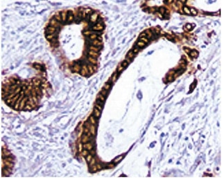
Immunocytochemistry: The HL3 antibody is recommended to test for immunohistochemical staining of acetone-fixed frozen sections. Tissues tested were mouse spleen and thymus. The clone HL3 is not recommended for zinc-fixed and formalin-fixed paraffin embedded sections. The antibody stains the dendritic cells and NK cells. The isotype control recommended for use with this antibody is purified hamster IgG (Cat. No. 553951). For optimal indirect immunohistochemical staining, the HL3 antibody should be titrated (1:10 to 1:50 dilution) and visualized via a three-step staining procedure in combination with biotinylated anti-hamster cocktail (Cat. No. 550335) as the secondary antibody and Streptavidin-HRP (Cat. No. 550946) together with the DAB dectection system (Cat. No. 550880). More conveniently, the Anti-Hamster Ig HRP detection kit (Cat. No. 551012) that contains the biotinylated secondary antibody, antibody diluent, streptavidin-HRP and DAB substrate can be used for staining. A detailed protocol of the immunohistochemical procedure is available at our website, http://www.bdbiosciences.com/support/resources.

Product Notices

  1. Since applications vary, each investigator should titrate the reagent to obtain optimal results.
  2. An isotype control should be used at the same concentration as the antibody of interest.
  3. Source of all serum proteins is from USDA inspected abattoirs located in the United States.
  4. Caution: Sodium azide yields highly toxic hydrazoic acid under acidic conditions. Dilute azide compounds in running water before discarding to avoid accumulation of potentially explosive deposits in plumbing.
  5. Although hamster immunoglobulin isotypes have not been well defined, BD Biosciences Pharmingen has grouped Armenian and Syrian hamster IgG monoclonal antibodies according to their reactivity with a panel of mouse anti-hamster IgG mAbs. A table of the hamster IgG groups, Reactivity of Mouse Anti-Hamster Ig mAbs, may be viewed at http://www.bdbiosciences.com/documents/hamster_chart_11x17.pdf.
  6. This antibody has been developed for the immunohistochemistry application. However, a routine immunohistochemistry test is not performed on every lot. Researchers are encouraged to titrate the reagent for optimal performance.
  7. Sodium azide is a reversible inhibitor of oxidative metabolism; therefore, antibody preparations containing this preservative agent must not be used in cell cultures nor injected into animals. Sodium azide may be removed by washing stained cells or plate-bound antibody or dialyzing soluble antibody in sodium azide-free buffer. Since endotoxin may also affect the results of functional studies, we recommend the NA/LE (No Azide/Low Endotoxin) antibody format, if available, for in vitro and in vivo use.
  8. Please refer to www.bdbiosciences.com/us/s/resources for technical protocols.
550283 Rev. 6
抗体の詳細
Down Arrow Up Arrow
HL3

The HL3 monoclonal antibody specifically binds to the integrin αx chain of gp150, 95 (CD11c/CD18). CD11c is expressed on dendritic cells, CD4- CD8+ intestinal intraepithelial lymphocytes (IEL) and some NK cells. It is upregulated on IEL and lymph-node T cells following in vivo activation. Cells of the monocyte/macrophage lineage have been reported to express low levels of CD11c. CD11c plays a role in binding of iC3b.

550283 Rev. 6
フォーマットの詳細
Down Arrow Up Arrow
Purified
Tissue culture supernatant is purified by either protein A/G or affinity purification methods. Both methods yield antibody in solution that is free of most other soluble proteins, lipids, etc. This format provides pure antibody that is suitable for a number of downstream applications including: secondary labeling for flow cytometry or microscopy, ELISA, Western blot, etc.
Purified
550283 Rev.6
引用&参考文献
Down Arrow Up Arrow

Development References (6)

  1. Burt BM, Plitas G, Stableford JA, et al. CD11c identifies a subset of murine liver natural killer cells that responds to adenoviral hepatitis. J Leukoc Biol. 2008; 84(4):1039-1046. (Clone-specific). View Reference
  2. Fagarasan S, Muramatsu M, Suzuki K, Nagaoka H, Hiai H, Honjo T. Critical roles of activation-induced cytidine deaminase in the homeostasis of gut flora. Science. 2002; 298(5597):1424-1427. (Clone-specific: Immunofluorescence). View Reference
  3. Huleatt JW, Lefrançois L. Antigen-driven induction of CD11c on intestinal intraepithelial lymphocytes and CD8+ T cells in vivo.. J Immunol. 1995; 154(11):5684-93. (Immunogen: Immunoprecipitation). View Reference
  4. Larson RS, Springer TA. Structure and function of leukocyte integrins. Immunol Rev. 1990; 114:181-217. (Biology). View Reference
  5. Metlay JP, Witmer-Pack MD, Agger R, Crowley MT, Lawless D, Steinman RM. The distinct leukocyte integrins of mouse spleen dendritic cells as identified with new hamster monoclonal antibodies. J Exp Med. 1990; 171(5):1753-1771. (Biology). View Reference
  6. Pulendran B, Lingappa J, Kennedy MK, et al. Developmental pathways of dendritic cells in vivo: distinct function, phenotype, and localization of dendritic cell subsets in FLT3 ligand-treated mice. J Immunol. 1997; 159(5):2222-2231. (Clone-specific: Immunohistochemistry). View Reference
すべて表示する (6) 表示項目を減らす
550283 Rev. 6

Please refer to Support Documents for Quality Certificates


Global - Refer to manufacturer's instructions for use and related User Manuals and Technical data sheets before using this products as described


Comparisons, where applicable, are made against older BD Technology, manual methods or are general performance claims.  Comparisons are not made against non-BD technologies, unless otherwise noted.

For Research Use Only. Not for use in diagnostic or therapeutic procedures.