-
抗体試薬
- フローサイトメトリー用試薬
-
ウェスタンブロッティング抗体試薬
- イムノアッセイ試薬
-
シングルセル試薬
- BD® AbSeq Assay
- BD Rhapsody™ Accessory Kits
- BD® OMICS-One Immune Profiler Protein Panel
- BD® Single-Cell Multiplexing Kit
- BD Rhapsody™ TCR/BCR Next Multiomic Assays
- BD Rhapsody™ Targeted mRNA Kits
- BD Rhapsody™ Whole Transcriptome Analysis (WTA) Amplification Kit
- BD® OMICS-Guard Sample Preservation Buffer
- BD Rhapsody™ ATAC-Seq Assays
- BD® OMICS-One Protein Panels
-
細胞機能評価のための試薬
-
顕微鏡・イメージング用試薬
-
細胞調製・分離試薬
-
- BD® AbSeq Assay
- BD Rhapsody™ Accessory Kits
- BD® OMICS-One Immune Profiler Protein Panel
- BD® Single-Cell Multiplexing Kit
- BD Rhapsody™ TCR/BCR Next Multiomic Assays
- BD Rhapsody™ Targeted mRNA Kits
- BD Rhapsody™ Whole Transcriptome Analysis (WTA) Amplification Kit
- BD® OMICS-Guard Sample Preservation Buffer
- BD Rhapsody™ ATAC-Seq Assays
- BD® OMICS-One Protein Panels
- Japan (Japanese)
-
Change country/language
Old Browser
Looks like you're visiting us from United States.
Would you like to stay on the current country site or be switched to your country?
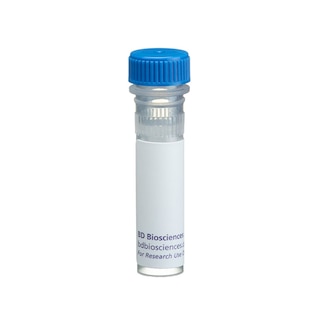
BD Pharmingen™ Purified Hamster Anti-Mouse CD3e
クローン 500A2 (RUO)


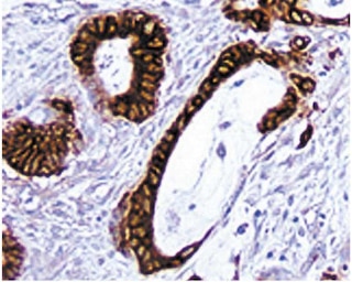
Immunohistochemical staining of T lymphocytes.
A frozen section from mouse spleen was stained with the Hamster Anti-Mouse CD3e(Cat. No. 550277) antibody. T lymphocytes can be identified by the intense brown labeling of their cell surface membranes (20X magnification).


BD Pharmingen™ Purified Hamster Anti-Mouse CD3e

Regulatory Statusの凡例
Any use of products other than the permitted use without the express written authorization of Becton, Dickinson and Company is strictly prohibited.
Preparation and Storage
推奨アッセイ手順
Immunohistochemistry: For optimal indirect immunohistochemical staining, this antibody should be titrated (e.g suggested starting dilutions of 1:10 to 1:50) and visualized via a three step staining procedure in combination with a Biotin Mouse Anti-Hamster IgG2 antibody (Cat. No. 554029) as the secondary antibody and Streptavidin-HRP (Cat. No. 550946) together with the DAB detection system (Cat. No. 550880).
Investigators should note that this antibody is not recommended for paraffin-embedded sections.
Product Notices
- Since applications vary, each investigator should titrate the reagent to obtain optimal results.
- Although hamster immunoglobulin isotypes have not been well defined, BD Biosciences Pharmingen has grouped Armenian and Syrian hamster IgG monoclonal antibodies according to their reactivity with a panel of mouse anti-hamster IgG mAbs. A table of the hamster IgG groups, Reactivity of Mouse Anti-Hamster Ig mAbs, may be viewed at http://www.bdbiosciences.com/documents/hamster_chart_11x17.pdf.
- Source of all serum proteins is from USDA inspected abattoirs located in the United States.
- Caution: Sodium azide yields highly toxic hydrazoic acid under acidic conditions. Dilute azide compounds in running water before discarding to avoid accumulation of potentially explosive deposits in plumbing.
- An isotype control should be used at the same concentration as the antibody of interest.
- This antibody has been developed for the immunohistochemistry application. However, a routine immunohistochemistry test is not performed on every lot. Researchers are encouraged to titrate the reagent for optimal performance.
- Please refer to www.bdbiosciences.com/us/s/resources for technical protocols.
関連製品


The 500A2 monoclonal antibody specifically binds to the 25-kDa ε chain of the T-cell receptor-associated CD3 complex expressed on mouse thymocytes, mature T lymphocytes, and NKT cells. Plate-bound and soluble forms of the 500A2 antibody can activate T cells in vitro. Activation of a mouse T-cell clone by the 500A2 antibody can be blocked by Fab fragments of the GK1.5 anti-CD4 antibody. This suggests that the 500A2 antibody may bind an epitope on CD3e close to a site at which CD4 associates with the T-cell receptor. The 500A2 antibody reportedly does not to crossreact with rat leukocytes.
Development References (3)
-
Allison JP, Havran WL, Poenie M, et al. Expression and function of CD3 on murine thymocytes. In: Kappler J, Davis M, ed. The T-Cell Receptor, UCLA Symposia, 73rd Edition. Los Angeles: 1988:33-45.
-
Havran WL, Poenie M, Kimura J, Tsien R, Weiss A, Allison JP. Expression and function of the CD3-antigen receptor on murine CD4+8+ thymocytes. Nature. 1987; 330(6144):170-173. (Biology: (Co)-stimulation, Immunohistochemistry). View Reference
-
Portoles P, Rojo J, Golby A, et al . Monoclonal antibodies to murine CD3 epsilon define distinct epitopes, one of which may interact with CD4 during T cell activation. J Immunol. 1989; 142(12):4169-4175. (Biology). View Reference
Please refer to Support Documents for Quality Certificates
Global - Refer to manufacturer's instructions for use and related User Manuals and Technical data sheets before using this products as described
Comparisons, where applicable, are made against older BD Technology, manual methods or are general performance claims. Comparisons are not made against non-BD technologies, unless otherwise noted.
For Research Use Only. Not for use in diagnostic or therapeutic procedures.