


製品詳細

BD Pharmingen™
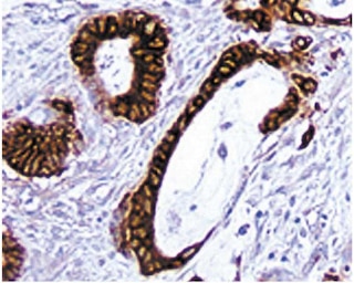
Goliath-related E3 ubiquitin-protein ligase 1, RNF128
Mouse (QC Testing)
Rat LOU, also known as Louvain, LOU/C, LOU/M IgG2a, κ
Mouse GRAIL (full-length) Recombinant Protein
Western blot (Routinely Tested), (Tested During Development)
62-66 kDa
0.5 mg/ml
AB_2869097
Aqueous buffered solution containing ≤0.09% sodium azide.
RUO
Preparation and Storage
The monoclonal antibody was purified from tissue culture supernatant or ascites by affinity chromatography. Store undiluted at 4°C.
Product Notices
- Since applications vary, each investigator should titrate the reagent to obtain optimal results.
- Caution: Sodium azide yields highly toxic hydrazoic acid under acidic conditions. Dilute azide compounds in running water before discarding to avoid accumulation of potentially explosive deposits in plumbing.
- Please refer to www.bdbiosciences.com/us/s/resources for technical protocols.
関連製品
Purified Rat IgG2a κ Isotype Control RUO
サイズ
0.25 mg
カタログ番号
559073
Biotin Goat Anti-Rat Ig RUO
サイズ
0.5 mg
カタログ番号
559286

Streptavidin HRP RUO
サイズ
50 mL
カタログ番号
550946

DAB Substrate Kit RUO
サイズ
500 Tests
カタログ番号
550880
557799 Rev. 7
Please refer to Support Documents for Quality Certificates
Global - Refer to manufacturer's instructions for use and related User Manuals and Technical data sheets before using this products as described
Comparisons, where applicable, are made against older BD Technology, manual methods or are general performance claims. Comparisons are not made against non-BD technologies, unless otherwise noted.
For Research Use Only. Not for use in diagnostic or therapeutic procedures.