


製品詳細

BD IMag™
CD3E; T3E; TCRE; cd 3; cd-3; cd3; CD3-epsilon; 916
Human (QC Testing)
Mouse IgG2a, κ
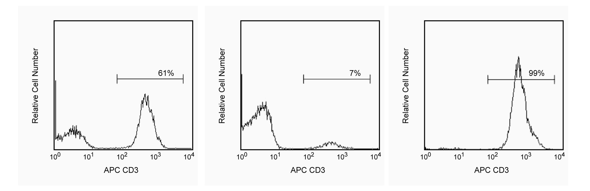
Cell separation (Routinely Tested)
V 5T-CD03.05
AB_398523
Aqueous buffered solution containing BSA and ≤0.09% sodium azide.
RUO
Preparation and Storage
Store undiluted at 4°C. The monoclonal antibody was purified from tissue culture supernatant or ascites by affinity chromatography. Antibody or streptavidin was conjugated to the magnetic particles under optimum conditions, and unconjugated antibody/streptavidin was removed.
推奨アッセイ手順
Product Notices
- BD IMag™ particles are prepared from carboxy-functionalized magnetic particles which are manufactured by Skold Technology and are licensed under US patent number 7,169,618.
- Ficoll-Paque is a trademark of Amersham Biosciences Limited.
- Caution: Sodium azide yields highly toxic hydrazoic acid under acidic conditions. Dilute azide compounds in running water before discarding to avoid accumulation of potentially explosive deposits in plumbing.
- Source of all serum proteins is from USDA inspected abattoirs located in the United States.
- Please refer to www.bdbiosciences.com/us/s/resources for technical protocols.
関連製品
Buffer (10X) RUO
サイズ
100 mL
カタログ番号
552362
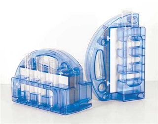
Cell Separation Magnet RUO
カタログ番号
552311
APC Mouse Anti-Human CD3 RUO
サイズ
100 Tests
カタログ番号
555335
552593 Rev. 8
Please refer to Support Documents for Quality Certificates
Global - Refer to manufacturer's instructions for use and related User Manuals and Technical data sheets before using this products as described
Comparisons, where applicable, are made against older BD Technology, manual methods or are general performance claims. Comparisons are not made against non-BD technologies, unless otherwise noted.
For Research Use Only. Not for use in diagnostic or therapeutic procedures.