Using the latest advances in flow cytometry to tackle challenges in extracellular vesicle characterization
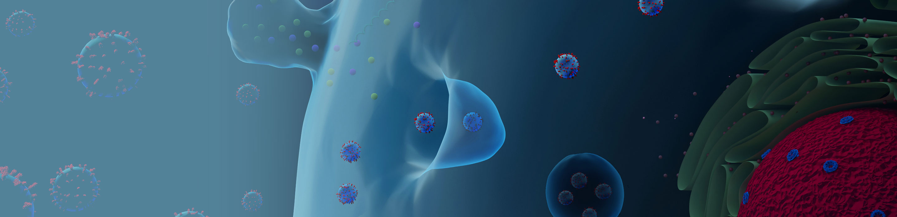
Extracellular vesicles (EVs), the small, heterogenous membrane-bound secretory particles that are released from myriad cell types, have gained prominence in recent years. Present in almost every bodily fluid, these apparently simple vesicles are quite complex in terms of their cargo (which can include nucleic acids, proteins and lipids) and their biologically active surface molecules that can be used as biomarkers as well as indicators of disease progression. The potential use of EVs as biomarkers in monitoring disease progression,1 as therapeutic targets for cancer,2 and as drug delivery systems3 has been investigated extensively.
Flow cytometry is the most commonly used analytical technique for EV characterization,4 but there are several challenges associated with this method. One of the challenges with using flow cytometry for EV characterization is the small size of EVs (which ranges from 30 to 1,000 nm)4 versus the typical range of detection of regular flow cytometers (10–100 µm). This, compounded by the composition of EVs and differences in their concentrations, makes characterization of EVs difficult. But advances in flow cytometry instrumentation in recent years has removed some of these barriers and shown promise for an explosion of growth in EV research. (See Nature article “Flow cytometry zooms in on extracellular vesicles” for more on this.)
For example, an advanced flow cytometer like the BD FACSymphony™ A1 Cell Analyzer with BD® Small Particle Detector provides a dedicated high-sensitivity detector for a side scatter channel (SP SSC) designed to resolve scatter of small particles such as EVs, viral particle, exosomes and others. This offers the ability to detect particles as small as 90-nm polystyrene beads without changing the instrument setup. In addition, new software plugins, such as the FlowJo™ Rosetta Calibration Plugin, enable utilizing the flow cytometry standard (FCS) data captured from Rosetta Calibration beads to derive calibrated size measurement of small particles.
EV researchers have utilized these new advances in innovative ways to resolve several issues that could not be addressed before. BD Biosciences, in collaboration with Nature, organized a webinar on the latest developments in extracellular vesicle analysis using flow cytometry with prominent EV researchers Dr. Jonni Moore of Penn Cytomics and Cell Sorting Resource Laboratory, Dr. Terry K. Morgan of Oregon Health & Science University and Dr. Edwin van der Pol of Amsterdam University Medical Centers.
In this webinar, Dr. Moore discusses how innovation in EV cytometry has helped her lab in mining of EV information from liquid biopsy samples. Dr. Morgan describes how he detected gestational phenotype differences using cell-specific EVs. Dr. Edwin van der Pol demonstrates how he used flow cytometry to measure concentrations of EVs that were <100 nm in a reproducible and scalable way and how EV flow cytometry could become the state-of-the art nanoparticle characterization technique.
Learn more about the BD FACSymphony™ A1 Cell Analyzer with BD® Small Particle Detector and see how it can be used in your EV research.
Watch the on-demand video entitled:
“Innovation in extracellular vesicle research with flow cytometry”.