





.png)


Preparation and Storage


Product Notices
- This reagent has been pre-diluted for use at the recommended Volume per Test. We typically use 1 × 10^6 cells in a 100-µl experimental sample (a test).
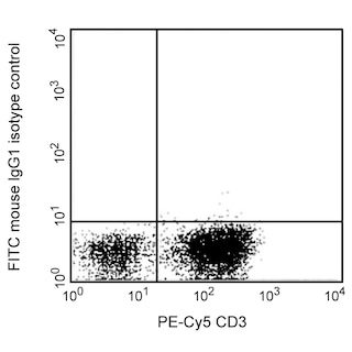
- An isotype control should be used at the same concentration as the antibody of interest.
- Source of all serum proteins is from USDA inspected abattoirs located in the United States.
- Caution: Sodium azide yields highly toxic hydrazoic acid under acidic conditions. Dilute azide compounds in running water before discarding to avoid accumulation of potentially explosive deposits in plumbing.
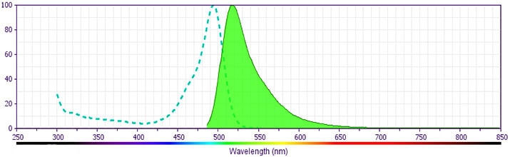
- For fluorochrome spectra and suitable instrument settings, please refer to our Multicolor Flow Cytometry web page at www.bdbiosciences.com/colors.
- Please refer to www.bdbiosciences.com/us/s/resources for technical protocols.
The P1H12 antibody reacts with a 118 kDa transmembrane glycoprotein also known as MCAM, MUC18, or Mel-CAM. CD146 is a member of the immunoglobulin superfamily and is expressed by angioblasts and mesenchymal stems cells and is strongly expressed by blood vessel endothelium and smooth muscle. CD146 is also expressed by melanoma cells, intermediate trophoblasts and a subpopulation of activated T cells. The P1H12 monoclonal antibody has been reported to block endothelial cell adhesion that is observed very early in embryogenesis.
Development References (7)
-
Elshal MF, Khan SS, Takahashi Y, Solomon MA, McCoy JP, Jr. CD146 (Mel-CAM), an adhesion marker of endothelial cells, is a novel marker of lymphocyte subset activation in normal peripheral blood. Blood. 2005; 106(8):2923-2924. (Clone-specific: Flow cytometry). View Reference
-
Kishimoto T. Tadamitsu Kishimoto .. et al., ed. Leucocyte typing VI : white cell differentiation antigens : proceedings of the sixth international workshop and conference held in Kobe, Japan, 10-14 November 1996. New York: Garland Pub.; 1997.
-
Sers C, Kirsch K, Rothbächer U, Riethmüller G, Johnson JP. Genomic organization of the melanoma-associated glycoprotein MUC18: implications for the evolution of the immunoglobulin domains. Proc Natl Acad Sci U S A. 1993; 90(18):8514-8518. (Biology). View Reference
-
Shih IM, Elder DE, Hsu MY, Herlyn M. Regulation of Mel-CAM/MUC18 expression on melanocytes of different stages of tumor progression by normal keratinocytes. Am J Pathol. 1994; 145(4):837-845. (Biology). View Reference
-
Shih IM. The role of CD146 (Mel-CAM) in biology and pathology. J Pathol. 1999; 189(1):4-11. (Biology). View Reference
-
Solovey A, Lin Y, Browne P, Choong S, Wayner E, Hebbel R P. Circulating activated endothelial cells in sickle cell anemia. N Engl J Med. 1997; 337(22):1584-1590. (Clone-specific: Fluorescence microscopy). View Reference
-
Solovey AN, Gui L, Chang L, Enenstein J, Browne PV, Hebbel RP. Identification and functional assessment of endothelial P1H12. J Lab Clin Med. 2001; 138(5):322-331. (Clone-specific). View Reference
Please refer to Support Documents for Quality Certificates
Global - Refer to manufacturer's instructions for use and related User Manuals and Technical data sheets before using this products as described
Comparisons, where applicable, are made against older BD Technology, manual methods or are general performance claims. Comparisons are not made against non-BD technologies, unless otherwise noted.
For Research Use Only. Not for use in diagnostic or therapeutic procedures.
