Preparation and Storage
推奨アッセイ手順
BD® CompBeads can be used as surrogates to assess fluorescence spillover (compensation). When fluorochrome conjugated antibodies are bound to BD® CompBeads, they have spectral properties very similar to cells. However, for some fluorochromes there can be small differences in spectral emissions compared to cells, resulting in spillover values that differ when compared to biological controls. It is strongly recommended that when using a reagent for the first time, users compare the spillover on cells and BD® CompBeads to ensure that BD® CompBeads are appropriate for your specific cellular application.
Product Notices
- Please refer to www.bdbiosciences.com/us/s/resources for technical protocols.
- Since applications vary, each investigator should titrate the reagent to obtain optimal results.
- An isotype control should be used at the same concentration as the antibody of interest.
- Caution: Sodium azide yields highly toxic hydrazoic acid under acidic conditions. Dilute azide compounds in running water before discarding to avoid accumulation of potentially explosive deposits in plumbing.
- This product is provided under an Agreement between BIOTIUM and BD Biosciences. This product, and only in the amount purchased by buyer, may be used solely for buyer’s own internal research, in a manner consistent with the accompanying product literature. No other right to use, sell or otherwise transfer (a) this product, or (b) its components is hereby granted expressly, by implication or by estoppel. This product is for research use only. Diagnostic uses require a separate license from Biotium, Inc. For information on purchasing a license to this product including for purposes other than research, contact Biotium, Inc., 3159 Corporate Place, Hayward, CA 94545, Tel: (510) 265-1027. Fax: (510) 265-1352. Email: btinfo@biotium.com.
- Please refer to http://regdocs.bd.com to access safety data sheets (SDS).
- Alexa Fluor™ is a trademark of Life Technologies Corporation.
- For U.S. patents that may apply, see bd.com/patents.
関連製品


.png?imwidth=320)
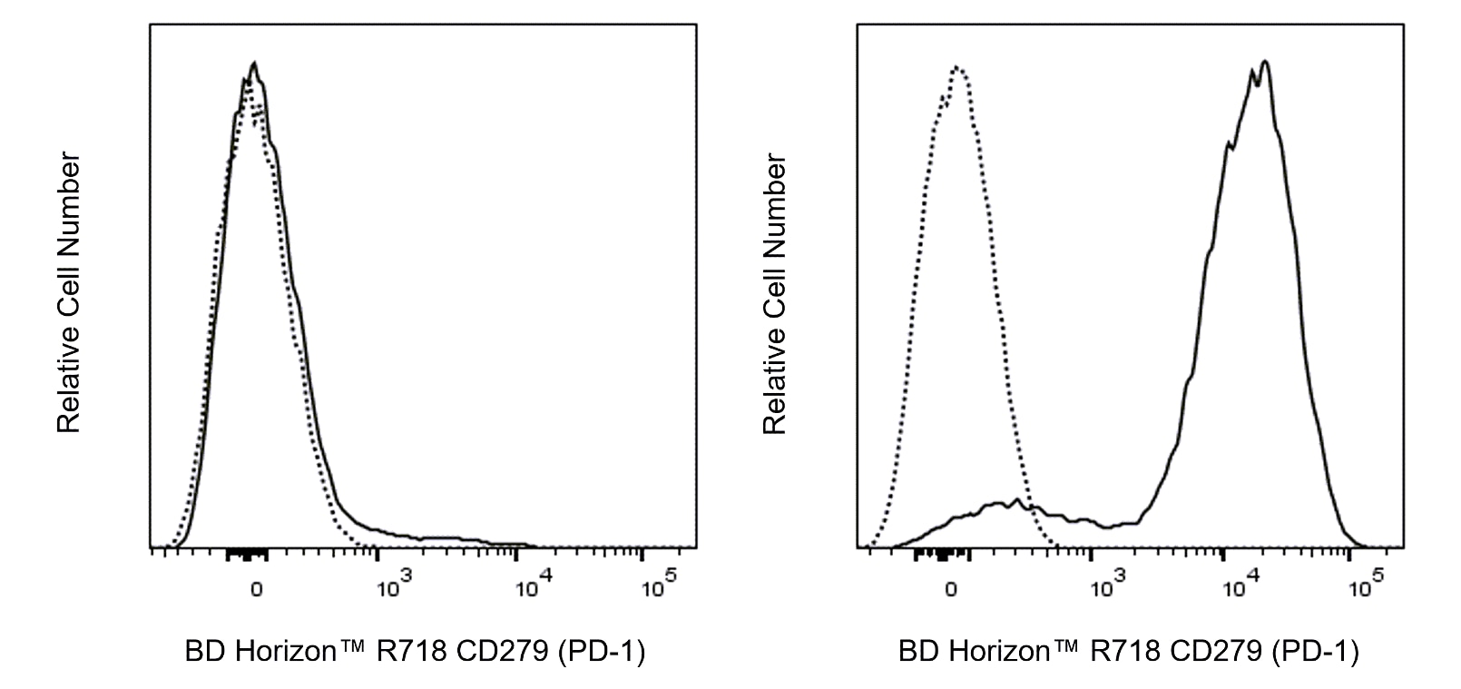
The 29F.1A12 monoclonal antibody specifically recognizes CD279 which is also known as Programmed Death-1 (PD-1). CD279 (PD-1) is a ~55-kDa type I transmembrane glycoprotein that is encoded by Pdcd1 (Programmed cell death 1) which belongs to the CD28/CTLA-4 family of immunoreceptors within the Ig superfamily. CD279 (PD-1) is comprised of an extracellular region with an IgV-like domain, a transmembrane sequence, and an intracellular region with an immunoreceptor tyrosine-based inhibitory motif (ITIM) and an immunoreceptor tyrosine-based switch motif (ITSM) that are associated with suppressive immunoregulatory functions. CD279 (PD-1) is variably expressed on some thymocyte subsets and developing B lymphocytes at the pro-B-cell stage. It is also inducibly expressed on activated myeloid cells, B cells, and T cells including exhausted T cells found in mice during chronic viral infections or cancer. Although this co-inhibitory receptor plays roles in mediating immunological tolerance and preventing autoimmune responses it can also inhibit protective immune responses against microbial infections and cancer. CD273 (also known as PD-L2 or B7-DC) and CD274 (PD-L1 or B7-H1) are members of the B7 family within the Ig superfamily. These molecules serve as ligands for CD279 (PD-1) and are variably expressed on lymphoid and nonlymphoid cell types including antigen-presenting cells and tumor cells. The 29F.1A12 antibody can reportedly block the binding of these ligands as well as other mouse PD-1-specific antibodies including clones J43, G4, and RMP1-14. Antibody-mediated inhibition of the interaction between PD-1 and its ligands can serve as an immune checkpoint blockade that can augment T-cell responses against tumor cells.

Development References (5)
-
Bu MT, Yuan L, Klee AN, Freeman GJ. A Comparison of Murine PD-1 and PD-L1 Monoclonal Antibodies.. Monoclon Antib Immunodiagn Immunother. 2022; 41(4):202-209. (Clone-specific: Blocking, Flow cytometry). View Reference
-
Liang SC, Latchman YE, Buhlmann JE, et al. Regulation of PD-1, PD-L1, and PD-L2 expression during normal and autoimmune responses.. Eur J Immunol. 2003; 33(10):2706-16. (Immunogen: Flow cytometry, Immunofluorescence, Immunohistochemistry). View Reference
-
Lázár-Molnár E, Gácser A, Freeman GJ, Almo SC, Nathenson SG, Nosanchuk JD. The PD-1/PD-L costimulatory pathway critically affects host resistance to the pathogenic fungus Histoplasma capsulatum.. Proc Natl Acad Sci U S A. 2008; 105(7):2658-63. (Clone-specific: In vivo exacerbation). View Reference
-
Polesso F, Munks MW, Rott KH, Smart S, Hill AB, Moran AE. PD-1-specific "Blocking" antibodies that deplete PD-1+ T cells present an inconvenient variable in preclinical immunotherapy experiments.. Eur J Immunol. 2021; 51(6):1473-1481. (Clone-specific). View Reference
-
Puzey MS, Craig CJ. Hydrometrocolpos--a case report.. S Afr Med J. 1992; 81(6):336-7. (Biology). View Reference
Please refer to Support Documents for Quality Certificates
Global - Refer to manufacturer's instructions for use and related User Manuals and Technical data sheets before using this products as described
Comparisons, where applicable, are made against older BD Technology, manual methods or are general performance claims. Comparisons are not made against non-BD technologies, unless otherwise noted.
For Research Use Only. Not for use in diagnostic or therapeutic procedures.