
Preparation and Storage
推奨アッセイ手順
BD™ CompBeads can be used as surrogates to assess fluorescence spillover (Compensation). When fluorochrome conjugated antibodies are bound to CompBeads, they have spectral properties very similar to cells. However, for some fluorochromes there can be small differences in spectral emissions compared to cells, resulting in spillover values that differ when compared to biological controls. It is strongly recommended that when using a reagent for the first time, users compare the spillover on cells and CompBead to ensure that BD Comp beads are appropriate for your specific cellular application.
For optimal and reproducible results, BD Horizon Brilliant Stain Buffer should be used anytime two or more BD Horizon Brilliant dyes are used in the same experiment. Fluorescent dye interactions may cause staining artifacts which may affect data interpretation. The BD Horizon Brilliant Stain Buffer was designed to minimize these interactions. More information can be found in the Technical Data Sheet of the BD Horizon Brilliant Stain Buffer (Cat. No. 563794/566349) or the BD Horizon Brilliant Stain Buffer Plus (Cat. No. 566385).
For optimal results, it is recommended to perform two washes after staining with antibodies. Cells may be prepared, stained with antibodies and washed twice with wash buffer per established protocols for immunofluorescent staining prior to acquisition on a flow cytometer. Performing fewer than the recommended wash steps may lead to increased spread of the negative population.
Product Notices
- Since applications vary, each investigator should titrate the reagent to obtain optimal results.
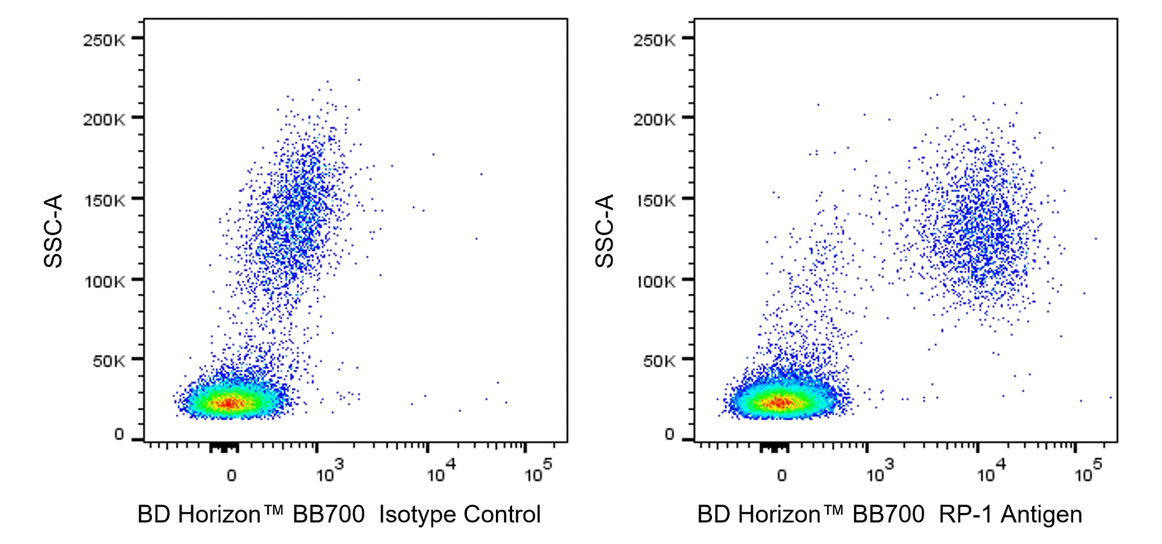
- An isotype control should be used at the same concentration as the antibody of interest.
- Caution: Sodium azide yields highly toxic hydrazoic acid under acidic conditions. Dilute azide compounds in running water before discarding to avoid accumulation of potentially explosive deposits in plumbing.
- For fluorochrome spectra and suitable instrument settings, please refer to our Multicolor Flow Cytometry web page at www.bdbiosciences.com/colors.
- BD Horizon Brilliant Stain Buffer is covered by one or more of the following US patents: 8,110,673; 8,158,444; 8,575,303; 8,354,239.
- BD Horizon Brilliant Blue 700 is covered by one or more of the following US patents: 8,455,613 and 8,575,303.
- Cy is a trademark of GE Healthcare.
- Please refer to http://regdocs.bd.com to access safety data sheets (SDS).
- Please refer to www.bdbiosciences.com/us/s/resources for technical protocols.
関連製品


The RP-1 monoclonal antibody specifically recognizes the RP-1 Antigen. This cell surface marker is expressed on rat peritoneal and peripheral blood neutrophils. Amongst bone marrow cells, the RP-1 Antigen is expressed on band form and mature neutrophils but is not expressed on promyelocytes, myelocytes, and metamyelocytes. The RP-1 antibody does not bind to either rat monocytes, macrophages, eosinophils or to peritoneal neutrophils from mice, rabbits, guinea pigs, or to human peripheral blood neutrophils. Expression of the RP-1 Antigen on rat peritoneal neutrophils is enhanced by cellular stimulation with Phorbol 12-Myristate 13-Acetate (PMA) or Concanavalin A (ConA). Immunoprecipitation and SDS-PAGE analysis of non-treated and PMA-activated rat neutrophil membranes with the RP-1 antibody revealed two main bands of approximately 85 kDa. The RP-1 antibody is also known as the Mouse Anti-Rat Granulocytes antibody.
The antibody was conjugated to BD Horizon BB700, which is part of the BD Horizon Brilliant™ Blue family of dyes. It is a polymer-based tandem dye developed exclusively by BD Biosciences. With an excitation max of 485 nm and an emission max of 693 nm, BD Horizon BB700 can be excited by the 488 nm laser and detected in a standard PerCP-Cy™5.5 set (eg, 695/40-nm filter). This dye provides a much brighter alternative to PerCP-Cy5.5 with less cross laser excitation off the 405 nm and 355 nm lasers.

Development References (3)
-
Francis WR, Ireland RE, Spear AM, et al. Flow Cytometric Analysis of Hematopoietic Populations in Rat Bone Marrow. Impact of Trauma and Hemorrhagic Shock.. Cytometry A. 2019; 95(11):1167-1177. (Clone-specific: Flow cytometry, Fluorescence activated cell sorting). View Reference
-
Gotoh S, Itoh M, Fujii Y, Arai S, Sendo F. Enhancement of the expression of a rat neutrophil-specific cell surface antigen by activation with phorbol myristate acetate and concanavalin A. J Immunol. 1986; 137(2):643-650. (Immunogen: Flow cytometry, Immunoprecipitation, Radioimmunoassay). View Reference
-
Skrajnar S, Anzur Lasnik M, Bedina Zavec A. A flow cytometric method for determination of the blood neutrophil fraction in rats.. J Am Assoc Lab Anim Sci. 2009; 48(2):152-6. (Clone-specific: Flow cytometry). View Reference
Please refer to Support Documents for Quality Certificates
Global - Refer to manufacturer's instructions for use and related User Manuals and Technical data sheets before using this products as described
Comparisons, where applicable, are made against older BD Technology, manual methods or are general performance claims. Comparisons are not made against non-BD technologies, unless otherwise noted.
For Research Use Only. Not for use in diagnostic or therapeutic procedures.