

Preparation and Storage
推奨アッセイ手順
Immunofluorescent Staining and Flow Cytometric Analysis: The staining technique and blocking controls are described in detail by C. Prussin and D. Metcalfe. Asuitable mouse IgG2b isotype control for assessing the level of background staining on human cells is recommended: use at comparable concentrations to antibody of interest (e.g., ≤ 0.125 µg Ab/1 million cells).
Product Notices
- Since applications vary, each investigator should titrate the reagent to obtain optimal results.
- An isotype control should be used at the same concentration as the antibody of interest.
- Caution: Sodium azide yields highly toxic hydrazoic acid under acidic conditions. Dilute azide compounds in running water before discarding to avoid accumulation of potentially explosive deposits in plumbing.
- For fluorochrome spectra and suitable instrument settings, please refer to our Multicolor Flow Cytometry web page at www.bdbiosciences.com/colors.
- Please refer to www.bdbiosciences.com/us/s/resources for technical protocols.
関連製品


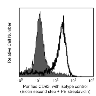
The R139 monoclonal antibody specifically binds to CD93 which is also known as Complement component C1q receptor (C1qR), C1q receptor 1 (C1qR1), or Matrix-remodeling-associated protein 4 (MXRA4). The immunogen used to generate the R139 hybridoma was a preparation of CD93 protein. Human CD93 is a transmembrane glycoprotein that is highly expressed on monocytes, macrophages, granulocytes, and endothelial cells but not on T and B lymphocytes. CD93 is also known as the C1q/MBL/SPA Receptor as it binds C1q, the recognition subunit of the first component (C1) of the complement pathway, as well as MBL (Mannose-binding-lectin) and SPA (Pulmonary Surfactant Protein A). Human C1qRp is involved in the C1q-mediated enhancement of phagocytosis. R139 is suitable to detect CD93 expression on cells of myeloid lineage by flow cytometry, and CD93 in cellular lysates by Western blotting or immunoprecipitation. In addition, R139 reportedly neutralizes C1q-mediated enhancement of phagocytosis. CD93 has also been reported to define a human stem cell population with hematopoietic and hepatic potential.

Development References (8)
-
Danet GH, Luongo JL, Butler G, et al. C1qRp defines a new human stem cell population with hematopoietic and hepatic potential.. Proc Natl Acad Sci USA. 2002; 99(16):10441-5. (Biology). View Reference
-
Guan E, Robinson SL, Goodman EB, Tenner AJ. Cell-surface protein identified on phagocytic cells modulates the C1q-mediated enhancement of phagocytosis. J Immunol. 1994; 152(8):4005-4016. (Biology). View Reference
-
Guan EN, Burgess WH, Robinson SL, Goodman EB, McTigue KJ, Tenner AJ. Phagocytic cell molecules that bind the collagen-like region of C1q. Involvement in the C1q-mediated enhancement of phagocytosis. J Biol Chem. 1991; 266(30):20345-20355. (Biology). View Reference
-
Nepomuceno RR, Henschen-Edman AH, Burgess WH, Tenner AJ. cDNA cloning and primary structure analysis of C1qR(P), the human C1q/MBL/SPA receptor that mediates enhanced phagocytosis in vitro. Immunity. 1997; 6(2):119-129. (Biology). View Reference
-
Nepomuceno RR, Ruiz S, Park M, Tenner AJ. C1qRP is a heavily O-glycosylated cell surface protein involved in the regulation of phagocytic activity. J Immunol. 1999; 162(6):3583-3589. (Biology). View Reference
-
Nepomuceno RR, Tenner AJ. C1qRP, the C1q receptor that enhances phagocytosis, is detected specifically in human cells of myeloid lineage, endothelial cells, and platelets. J Immunol. 1998; 160(4):1929-1935. (Biology). View Reference
-
Prussin C, Metcalfe DD. Detection of intracytoplasmic cytokine using flow cytometry and directly conjugated anti-cytokine antibodies. J Immunol Methods. 1995; 188(1):117-128. (Methodology). View Reference
-
Tenner AJ. C1q receptors: regulating specific functions of phagocytic cells. Immunobiology. 1998; 199(2):250-264. (Biology). View Reference
Please refer to Support Documents for Quality Certificates
Global - Refer to manufacturer's instructions for use and related User Manuals and Technical data sheets before using this products as described
Comparisons, where applicable, are made against older BD Technology, manual methods or are general performance claims. Comparisons are not made against non-BD technologies, unless otherwise noted.
For Research Use Only. Not for use in diagnostic or therapeutic procedures.