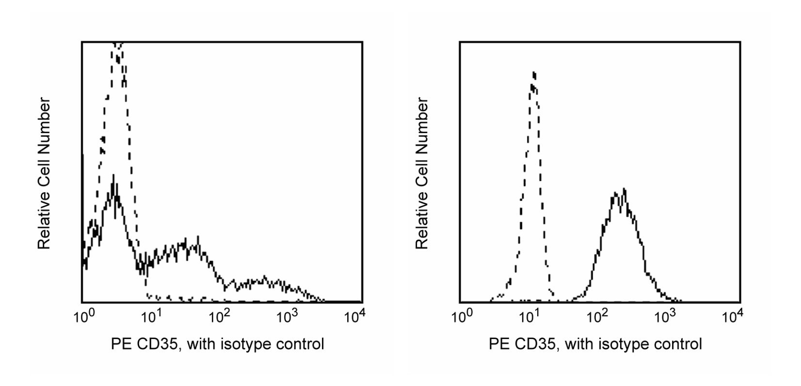
PE Mouse Anti-Human CD35
Clone E11 (RUO)
- Brand BD Pharmingen™
- Alternative Name CR1; Complement receptor type 1; C3b/C4b receptor; C3BR; C4BR; KN
- Vol. Per Test 20 µl
- Isotype Mouse IgG1, κ
- Reactivity Human (QC Testing)
- Application
Flow cytometry (Routinely Tested)
- Immunogen Human Cells of the Monocyte Lineage
- Workshop No. III 204
- Entrez Gene ID 1378
- Storage Buffer Aqueous buffered solution containing BSA and ≤0.09% sodium azide.
- Regulatory Status RUO
Regulatory Status Legend
Description
The E11 monoclonal antibody specifically binds to CD35. CD35 is also known as Complement receptor type 1 (CR1), C3b/C4b receptor, C3BR, C4BR, Immune adherence receptor, or KN. CD35 is a type I transmembrane glycoprotein that exists in four allelic forms of 160, 190, 220 and 250 kDa. CD35 serves as a receptor for complement fragments C3b, iC3b, C3dg, C4b, iC3, and iC4. It enhances phagocytosis by neutrophils and monocytes and regulates complement activation. It is expressed on erythrocytes, granulocytes, monocytes, B cells, and some dendritic cells, T cells, and NK cells. It binds complement components C3b and C4b, mediating. This antibody cannot inhibit the phagocytic capacity of granulocytes. The CD35 antibody is useful in studies of cells that express complement receptors.
Format
- Format PE
- Excitation Source Blue 488 nm,Green 532 nm,Yellow/Green 561 nm
- Excitation Max 496 nm
- Emission Max 578 nm
R-phycoerythrin (PE) is an accessory photosynthetic pigment found in red algae. It exists in vitro as a 240-kDa protein with 23 phycoerythrobilin chromophores per molecule. This makes PE one of the brightest fluorochromes for flow cytometry applications, but its photobleaching properties make it unsuitable for fluorescence microscopy.
Suggested Companion Products
PE Mouse IgG1, κ Isotype Control RUO
100 Tests
Cat No: 555749
Stain Buffer (FBS) RUO
500 mL
Cat No: 554656
Stain Buffer (BSA) RUO
500 mL
Cat No: 554657
Lysing Buffer RUO
100 mL
Cat No: 555899
Lysing Solution 10X Concentrate RUO (GMP)
100
Cat No: 349202
Resources & Tools | ||||||
---|---|---|---|---|---|---|
Spectrum Viewer | Panel Designer | Spectrum Viewer | Download TDS | Regulatory Document Website |
Preparation and Storage
Store undiluted at 4°C and protected from prolonged exposure to light. Do not freeze.
The monoclonal antibody was purified from tissue culture supernatant or ascites by affinity chromatography.
The antibody was conjugated with R-PE under optimum conditions, and unconjugated antibody and free PE were removed.
Product Notices
- This reagent has been pre-diluted for use at the recommended Volume per Test. We typically use 1 × 10^6 cells in a 100-µl experimental sample (a test).
- An isotype control should be used at the same concentration as the antibody of interest.
- Source of all serum proteins is from USDA inspected abattoirs located in the United States.
- Caution: Sodium azide yields highly toxic hydrazoic acid under acidic conditions. Dilute azide compounds in running water before discarding to avoid accumulation of potentially explosive deposits in plumbing.
- For fluorochrome spectra and suitable instrument settings, please refer to our Multicolor Flow Cytometry web page at www.bdbiosciences.com/colors.
- Please refer to www.bdbiosciences.com/us/s/resources for technical protocols.