-
抗体試薬
- フローサイトメトリー用試薬
-
ウェスタンブロッティング抗体試薬
- イムノアッセイ試薬
-
シングルセル試薬
- BD® AbSeq Assay | シングルセル試薬
- BD Rhapsody™ Accessory Kits | シングルセル試薬
- BD® Single-Cell Multiplexing Kit | シングルセル試薬
- BD Rhapsody™ Targeted mRNA Kits | シングルセル試薬
- BD Rhapsody™ Whole Transcriptome Analysis (WTA) Amplification Kit | シングルセル試薬
- BD Rhapsody™ TCR/BCR Profiling Assays (VDJ Assays) | シングルセル試薬
- BD® OMICS-Guard Sample Preservation Buffer
- BD Rhapsody™ ATAC-Seq Assays
-
細胞機能評価のための試薬
-
顕微鏡・イメージング用試薬
-
細胞調製・分離試薬
-
- BD® AbSeq Assay | シングルセル試薬
- BD Rhapsody™ Accessory Kits | シングルセル試薬
- BD® Single-Cell Multiplexing Kit | シングルセル試薬
- BD Rhapsody™ Targeted mRNA Kits | シングルセル試薬
- BD Rhapsody™ Whole Transcriptome Analysis (WTA) Amplification Kit | シングルセル試薬
- BD Rhapsody™ TCR/BCR Profiling Assays (VDJ Assays) | シングルセル試薬
- BD® OMICS-Guard Sample Preservation Buffer
- BD Rhapsody™ ATAC-Seq Assays
- Japan (Japanese)
-
Change country/language
Old Browser
Looks like you're visiting us from {countryName}.
Would you like to stay on the current country site or be switched to your country?


Western blot analysis of tomosyn on a rat cerebrum lysate (left). Lane 1: 1:250, Lane 2: 1:500, Lane 3: 1:1000 dilution of the anti- tomosyn antibody. Immunofluorescent staining of SK-N-SH cells (right). Cells were seeded in a 384 well collagen coated Microplates (Material # 353962) at ~ 8,000 cells per well. After overnight incubation, cells were stained using the Triton X100 fix/perm protocol (see Recommended Assay Procedure; Bioimaging protocol link) and the anti- Tomosyn antibody. The second step reagent was Alexa Fluor® 488 goat anti mouse Ig (Invitrogen)(pseudo colored green). Cell nuclei were counter stained with Hoechst 33342 (pseudo colored blue). The image was taken on a BD Pathway™ 855 or 435 Bioimager System using a 20x objective and merged using the BD AttoVison ™ software. This antibody also stained SH-SY5Y, SK-N-SH, C6, U87 and U373 cells using both the Triton X100 and methanol fix/perm protocols (see Recommended Assay Procedure; Bioimaging protocol link).


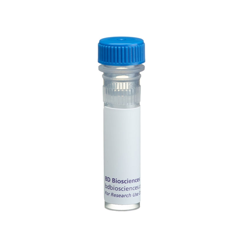
BD Transduction Laboratories™ Purified Mouse Anti-Tomosyn

Regulatory Statusの凡例
Any use of products other than the permitted use without the express written authorization of Becton, Dickinson and Company is strictly prohibited.
Preparation and Storage
Product Notices
- Since applications vary, each investigator should titrate the reagent to obtain optimal results.
- Caution: Sodium azide yields highly toxic hydrazoic acid under acidic conditions. Dilute azide compounds in running water before discarding to avoid accumulation of potentially explosive deposits in plumbing.
- Source of all serum proteins is from USDA inspected abattoirs located in the United States.
- This antibody has been developed and certified for the bioimaging application. However, a routine bioimaging test is not performed on every lot. Researchers are encouraged to titrate the reagent for optimal performance.
- Sodium azide is a reversible inhibitor of oxidative metabolism; therefore, antibody preparations containing this preservative agent must not be used in cell cultures nor injected into animals. Sodium azide may be removed by washing stained cells or plate-bound antibody or dialyzing soluble antibody in sodium azide-free buffer. Since endotoxin may also affect the results of functional studies, we recommend the NA/LE (No Azide/Low Endotoxin) antibody format, if available, for in vitro and in vivo use.
- Alexa Fluor® is a registered trademark of Molecular Probes, Inc., Eugene, OR.
- Triton is a trademark of the Dow Chemical Company.
- Please refer to www.bdbiosciences.com/us/s/resources for technical protocols.
関連製品

.png?imwidth=320)

Neuronal signal transmission and neurotransmitter release from the presynaptic nerve terminal is mediated by the synaptic vesicle cycle. Syntaxin plays a central role during vesicle docking and fusion through interactions with many vesicle components. During docking, vSNAREs (VAMP/synaptobrevin, synaptotagmin) on the synaptic vesicle and tSNAREs (SNAP-25, syntaxin) on the plasma membrane interact to form a 7S complex, which is essential to docking and fusion. Syntaxin associates with munc18/n-sec1 prior to and/or during the formation of the 7S complex. This interaction may inhibit syntaxin binding proteins (VAMP, SNAP-25) that facilitate vesicle docking or fusion. Tomosyn, a syntaxin binding protein, displaces munc18 from syntaxin-1 and forms a novel 10S complex with syntaxin-1, SNAP-25, and synaptogamin. There are two splice variants of tomosyn designated b-tomosyn and s-tomosyn, while the original is referred to as m-tomosyn. Although b-tomosyn is ubiquitously expressed, s-tomosyn and m-tomosyn are expressed primarily in brain.
Development References (2)
-
Fujita Y, Shirataki H, Sakisaka T, et al. Tomosyn: a syntaxin-1-binding protein that forms a novel complex in the neurotransmitter release process. Neuron. 1998; 20(5):905-915. (Biology). View Reference
-
Yokoyama S, Shirataki H, Sakisaka T, Takai Y. Three splicing variants of tomosyn and identification of their syntaxin-binding region. Biochem Biophys Res Commun. 1999; 256(1):218-222. (Biology). View Reference
Please refer to Support Documents for Quality Certificates
Global - Refer to manufacturer's instructions for use and related User Manuals and Technical data sheets before using this products as described
Comparisons, where applicable, are made against older BD Technology, manual methods or are general performance claims. Comparisons are not made against non-BD technologies, unless otherwise noted.
For Research Use Only. Not for use in diagnostic or therapeutic procedures.
Report a Site Issue
This form is intended to help us improve our website experience. For other support, please visit our Contact Us page.