

Preparation and Storage
推奨アッセイ手順
Immunohistochemistry: The MECA-32 antibody specific for mouse panendothelial cell antigen is recommended to test for immunohistochemical staining of acetone-fixed frozen sections. Tissues tested were mouse spleen, thymus and small intestine. The antibody stains endothelial cells. The isotype control recommended for use with this antibody is Purified Rat IgG2a κ Isotype Control (Cat. No. 559073). For optimal indirect immunohistochemical staining, the MECA-32 antibody should be titrated (1:10 to 1:50 dilution) and visualized via a three-step staining procedure in combination with Biotin Goat Anti-Rat Ig (Cat. No. 559286) as the secondary antibody and Streptavidin-HRP (Cat. No. 550946) together with the DAB detection system (Cat. No. 550880). A detailed protocol of the immunohistochemical procedure is available on our website, http://www.bdbiosciences.com/us/s/resources, under "Cell Biology (WB, IP, IHC, IF)".
Product Notices
- Since applications vary, each investigator should titrate the reagent to obtain optimal results.
- An isotype control should be used at the same concentration as the antibody of interest.
- Source of all serum proteins is from USDA inspected abattoirs located in the United States.
- Caution: Sodium azide yields highly toxic hydrazoic acid under acidic conditions. Dilute azide compounds in running water before discarding to avoid accumulation of potentially explosive deposits in plumbing.
- Sodium azide is a reversible inhibitor of oxidative metabolism; therefore, antibody preparations containing this preservative agent must not be used in cell cultures nor injected into animals. Sodium azide may be removed by washing stained cells or plate-bound antibody or dialyzing soluble antibody in sodium azide-free buffer. Since endotoxin may also affect the results of functional studies, we recommend the NA/LE (No Azide/Low Endotoxin) antibody format, if available, for in vitro and in vivo use.
- Please refer to www.bdbiosciences.com/us/s/resources for technical protocols.
関連製品


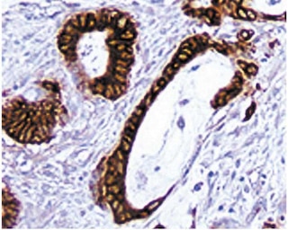


The MECA-32 antibody reacts with a dimer of 50-55–kDa subunits expressed on most or all endothelial cells in the embryonic and adult mouse, with the exception of cardiac and skeletal muscle and the brain. Normally in skeletal and cardiac muscle, MECA-32 antigen expression is limited to small arterioles and venules; however, under conditions of inflammation, it can be induced on previously non-expressing vessels in cardiac muscle. In the central nervous system (CNS), the panendothelial cell antigen expression is developmentally regulated. During embryonic development, the antigen is found on brain vasculature up to day 16 of gestation, after which it disappears. The cessation of MECA-32 antigen expression in the CNS may be associated with the establishment of the blood-brain barrier, which begins on day 16 of gestation. In the adult mouse, inflammation in the CNS can lead to re-expression of the panendothelial cell antigen.
Development References (5)
-
Bergese SD, Pelletier RP, Ohye RG, Vallera DA, Orosz CG. Treatment of mice with anti-CD3 mAb induces endothelial vascular cell adhesion molecule-1 expression. Transplantation. 1994; 57(5):711-717. (Biology). View Reference
-
Engelhardt B, Conley FK, Butcher EC. Cell adhesion molecules on vessels during inflammation in the mouse central nervous system. J Neuroimmunol. 1994; 51(2):199-208. (Biology). View Reference
-
Hallmann R, Mayer DN, Berg EL, Broermann R, Butcher EC. Novel mouse endothelial cell surface marker is suppressed during differentiation of the blood brain barrier. Dev Dyn. 1995; 202(4):325-332. (Biology). View Reference
-
Leppink DM, Bishop DK, Sedmak DD, et al. Inducible expression of an endothelial cell antigen on murine myocardial vasculature in association with interstitial cellular infiltration. Transplantation. 1989; 48(5):874-877. (Immunogen). View Reference
-
Orosz CG, van Buskirk A, Sedmak DD, Kincade P, Miyake K, Pelletier RP. Role of the endothelial adhesion molecule VCAM in murine cardiac allograft rejection. Immunol Lett. 1992; 32(1):7-12. (Biology). View Reference
Please refer to Support Documents for Quality Certificates
Global - Refer to manufacturer's instructions for use and related User Manuals and Technical data sheets before using this products as described
Comparisons, where applicable, are made against older BD Technology, manual methods or are general performance claims. Comparisons are not made against non-BD technologies, unless otherwise noted.
For Research Use Only. Not for use in diagnostic or therapeutic procedures.