-
抗体試薬
- フローサイトメトリー用試薬
-
ウェスタンブロッティング抗体試薬
- イムノアッセイ試薬
-
シングルセル試薬
- BD® AbSeq Assay | シングルセル試薬
- BD Rhapsody™ Accessory Kits | シングルセル試薬
- BD® Single-Cell Multiplexing Kit | シングルセル試薬
- BD Rhapsody™ Targeted mRNA Kits | シングルセル試薬
- BD Rhapsody™ Whole Transcriptome Analysis (WTA) Amplification Kit | シングルセル試薬
- BD Rhapsody™ TCR/BCR Profiling Assays (VDJ Assays) | シングルセル試薬
- BD® OMICS-Guard Sample Preservation Buffer
- BD Rhapsody™ ATAC-Seq Assays
-
細胞機能評価のための試薬
-
顕微鏡・イメージング用試薬
-
細胞調製・分離試薬
-
- BD® AbSeq Assay | シングルセル試薬
- BD Rhapsody™ Accessory Kits | シングルセル試薬
- BD® Single-Cell Multiplexing Kit | シングルセル試薬
- BD Rhapsody™ Targeted mRNA Kits | シングルセル試薬
- BD Rhapsody™ Whole Transcriptome Analysis (WTA) Amplification Kit | シングルセル試薬
- BD Rhapsody™ TCR/BCR Profiling Assays (VDJ Assays) | シングルセル試薬
- BD® OMICS-Guard Sample Preservation Buffer
- BD Rhapsody™ ATAC-Seq Assays
- Japan (Japanese)
-
Change country/language
Old Browser
Looks like you're visiting us from {countryName}.
Would you like to stay on the current country site or be switched to your country?


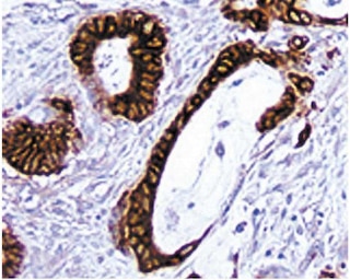
Immunohistochemical staining of CD11b positive cells. An acetone-fixed, frozen section of normal mouse spleen was stained with the Purified Rat Anti-CD11b antibody (Cat. No. 550282). Three-step staining was carried out with Biotin Goat Anti-Rat Ig (Cat. No. 559286) as the secondary antibody and Streptavidin HRP (Cat. No. 550946) together with a DAB detection system (Cat. No. 550880). CD11b positive cells can be identified by the intense brown labeling of cell surface membranes (20X magnification).


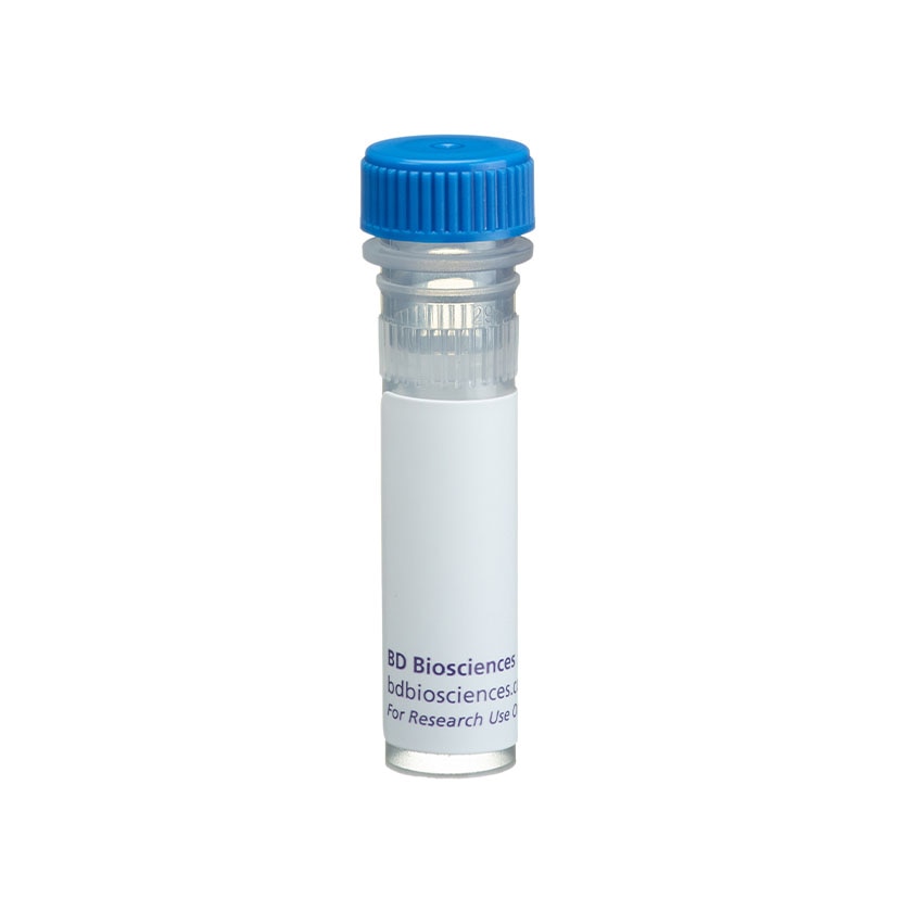
BD Pharmingen™ Purified Rat Anti-CD11b

Regulatory Statusの凡例
Any use of products other than the permitted use without the express written authorization of Becton, Dickinson and Company is strictly prohibited.
Preparation and Storage
推奨アッセイ手順
Immunohistochemistry: The Purified Rat Anti-Mouse CD11b antibody stains mouse macrophages, granulocytes, dendritic cells, and NK cells. For optimal indirect immunohistochemical staining, investigators are encouraged to titrate this antibody (suggested starting range of a 1:10 to 1:50 dilution) and visualize via a three-step staining procedure using a combination of Biotin Goat Anti-Rat Ig (Cat. No. 559286) as the secondary antibody and Streptavidin HRP (Cat. No. 550946) together with a DAB detection system (Cat. No. 550880). Investigators should note that this antibody is not recommended for formalin-fixed paraffin embedded sections.
Product Notices
- Since applications vary, each investigator should titrate the reagent to obtain optimal results.
- An isotype control should be used at the same concentration as the antibody of interest.
- Source of all serum proteins is from USDA inspected abattoirs located in the United States.
- Caution: Sodium azide yields highly toxic hydrazoic acid under acidic conditions. Dilute azide compounds in running water before discarding to avoid accumulation of potentially explosive deposits in plumbing.
- Sodium azide is a reversible inhibitor of oxidative metabolism; therefore, antibody preparations containing this preservative agent must not be used in cell cultures nor injected into animals. Sodium azide may be removed by washing stained cells or plate-bound antibody or dialyzing soluble antibody in sodium azide-free buffer. Since endotoxin may also affect the results of functional studies, we recommend the NA/LE (No Azide/Low Endotoxin) antibody format, if available, for in vitro and in vivo use.
- This antibody has been developed for the immunohistochemistry application. However, a routine immunohistochemistry test is not performed on every lot. Researchers are encouraged to titrate the reagent for optimal performance.
- Species cross-reactivity detected in product development may not have been confirmed on every format and/or application.
- Please refer to www.bdbiosciences.com/us/s/resources for technical protocols.
関連製品


The M1/70 monoclonal antibody specifically binds to CD11b, also known as Integrin alpha M (Itgam or αM). CD11b is a 170-kDa type 1 transmembrane glycoprotein and belongs to the Integrin alpha chain family. CD11b serves as the alpha chain of the heterodimeric Mac-1 integrin (CD11b/CD18, αMβ2), also known as complement receptor 3 (CR3). Mac-1 mediates adhesion to ICAM-1 (CD54), ICAM-2 (CD102), fibrinogen and binding to C3bi. Mac-1 is expressed at varying levels on granulocytes, macrophages, myeloid-derived dendritic cells, natural killer cells, microglia, and B-1 B lymphocytes. Mac-1 expression is rapidly upregulated on neutrophils after activation, in the same time period that CD62L (L-selectin) is shed from the cell surface. The M1/70 antibody reportedly blocks cell adherence and C3bi binding but does not block cell-mediated lysis. Cross-reaction of the M1/70 antibody with CD11b expressed on human monocytes, polymorphonuclear leukocytes, and NK cells has been reported.
Development References (8)
-
Ault KA, Springer TA. Cross-reaction of a rat-anti-mouse phagocyte-specific monoclonal antibody (anti-Mac-1) with human monocytes and natural killer cells. J Immunol. 1981; 126(1):359-364. (Biology). View Reference
-
Flotte TJ, Springer TA, Thorbecke GJ. Dendritic cell and macrophage staining by monoclonal antibodies in tissue sections and epidermal sheets. Am J Pathol. 1983; 111(1):112-124. (Biology). View Reference
-
Leenen PJ, de Bruijn MF, Voerman JS, Campbell PA, van Ewijk W. Markers of mouse macrophage development detected by monoclonal antibodies. J Immunol Methods. 1994; 174(1-2):5-19. (Biology). View Reference
-
Sanchez-Madrid F, Simon P, Thompson S, Springer TA. Mapping of antigenic and functional epitopes on the alpha- and beta-subunits of two related mouse glycoproteins involved in cell interactions, LFA-1 and Mac-1. J Exp Med. 1983; 158(2):586-602. (Biology: Blocking, Immunoprecipitation). View Reference
-
Springer T, Galfre G, Secher DS, Milstein C. Mac-1: a macrophage differentiation antigen identified by monoclonal antibody. Eur J Immunol. 1979; 9(4):301-306. (Biology: Immunoprecipitation). View Reference
-
Springer T, Galfre G, Secher DS, Milstein C. Monoclonal xenogeneic antibodies to murine cell surface antigens: identification of novel leukocyte differentiation antigens. Eur J Immunol. 1978; 8(8):539-551. (Immunogen: Immunoprecipitation). View Reference
-
Springer TA, Davignon D, Ho MK, Kurzinger K, Martz E, Sanchez-Madrid F. LFA-1 and Lyt-2,3, molecules associated with T lymphocyte-mediated killing; and Mac-1, an LFA-1 homologue associated with complement receptor function. Immunol Rev. 1982; 68:171-195. (Biology: Blocking). View Reference
-
Vremec D, Zorbas M, Scollay R, et al. The surface phenotype of dendritic cells purified from mouse thymus and spleen: investigation of the CD8 expression by a subpopulation of dendritic cells. J Exp Med. 1992; 176(1):47-58. (Biology). View Reference
Please refer to Support Documents for Quality Certificates
Global - Refer to manufacturer's instructions for use and related User Manuals and Technical data sheets before using this products as described
Comparisons, where applicable, are made against older BD Technology, manual methods or are general performance claims. Comparisons are not made against non-BD technologies, unless otherwise noted.
For Research Use Only. Not for use in diagnostic or therapeutic procedures.
Report a Site Issue
This form is intended to help us improve our website experience. For other support, please visit our Contact Us page.