-
抗体試薬
- フローサイトメトリー用試薬
-
ウェスタンブロッティング抗体試薬
- イムノアッセイ試薬
-
シングルセル試薬
- BD® AbSeq Assay | シングルセル試薬
- BD Rhapsody™ Accessory Kits | シングルセル試薬
- BD® Single-Cell Multiplexing Kit | シングルセル試薬
- BD Rhapsody™ Targeted mRNA Kits | シングルセル試薬
- BD Rhapsody™ Whole Transcriptome Analysis (WTA) Amplification Kit | シングルセル試薬
- BD® OMICS-Guard Sample Preservation Buffer
- BD Rhapsody™ ATAC-Seq Assays
- BD Rhapsody™ TCR/BCR Next Multiomic Assays
-
細胞機能評価のための試薬
-
顕微鏡・イメージング用試薬
-
細胞調製・分離試薬
-
- BD® AbSeq Assay | シングルセル試薬
- BD Rhapsody™ Accessory Kits | シングルセル試薬
- BD® Single-Cell Multiplexing Kit | シングルセル試薬
- BD Rhapsody™ Targeted mRNA Kits | シングルセル試薬
- BD Rhapsody™ Whole Transcriptome Analysis (WTA) Amplification Kit | シングルセル試薬
- BD® OMICS-Guard Sample Preservation Buffer
- BD Rhapsody™ ATAC-Seq Assays
- BD Rhapsody™ TCR/BCR Next Multiomic Assays
- Japan (Japanese)
-
Change country/language
Old Browser
Looks like you're visiting us from {countryName}.
Would you like to stay on the current country site or be switched to your country?


Left Figure: Western blot analysis of Nur77 expression on mouse thymocytes. Thymocytes were stimulated with PMA (20 ng/ml) and ionomycin (500 ng/ml at 37°C for 2 hr). Lysates were prepared and separated by SDS/PAGE. Blots were probed with Purified Mouse Anti-Nur77 (Cat. No. 554088) at concentrations of 2.0 (lane 1), 1.0 (lane 2), and 0.5 µg/ml (lane 3). Visualization was carried out with HRP Goat Anti-Mouse Ig (Cat. No. 554002). Nur77 is detected as a protein of ~77 kDa. Right Figure: Flow cytometric analysis of Nur77 expression on stimulated mouse thymocytes. Thymocytes from C57/BL/6 mice were left unstimulated (gray line histogram) or stimulated (black line histograms) with PMA (20ng/ml) and Ionomycin (500ng/ml) for 2 hours and then stained intracellularly with Purified Mouse IgG1, k Isotype Control (Cat.No. 554121; black dashed line) or Purified Mouse Anti-Nur77 antibody (black solid line) at 0.125ug/test using BD Pharmingen™ Transcription Factor Buffer Set (Cat. No. 562574). Purified antibody was followed by PE conjugated Goat Anti-Mouse Ig (Multiple Absorption Secondary antibody (Cat.No. 550589). Histogram plots were derived from gated events with the forward and side light-scatter characteristics of viable cells. Flow cytometric analysis was performed using a BD LSRFortessa™ X-20 Flow Cytometer System. Data shown on this Technical Data Sheet are not lot specific.


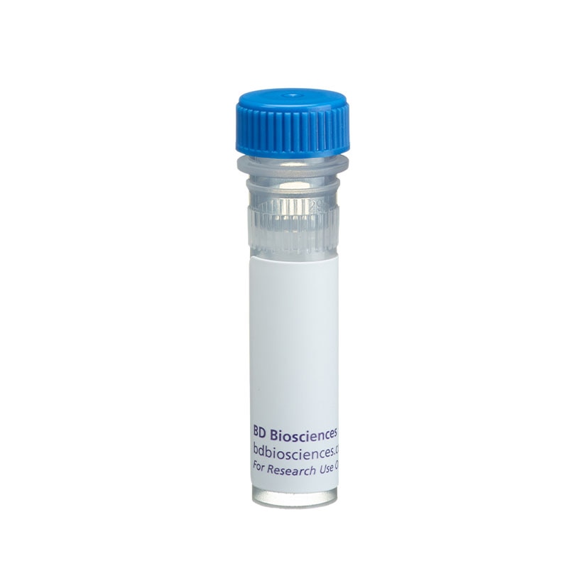
BD Pharmingen™ Purified Mouse Anti-Nur77

Regulatory Statusの凡例
Any use of products other than the permitted use without the express written authorization of Becton, Dickinson and Company is strictly prohibited.
Preparation and Storage
推奨アッセイ手順
Mouse thymocytes treated with PMA and ionomycin are suggested as a positive control.
Product Notices
- Since applications vary, each investigator should titrate the reagent to obtain optimal results.
- An isotype control should be used at the same concentration as the antibody of interest.
- Caution: Sodium azide yields highly toxic hydrazoic acid under acidic conditions. Dilute azide compounds in running water before discarding to avoid accumulation of potentially explosive deposits in plumbing.
- Sodium azide is a reversible inhibitor of oxidative metabolism; therefore, antibody preparations containing this preservative agent must not be used in cell cultures nor injected into animals. Sodium azide may be removed by washing stained cells or plate-bound antibody or dialyzing soluble antibody in sodium azide-free buffer. Since endotoxin may also affect the results of functional studies, we recommend the NA/LE (No Azide/Low Endotoxin) antibody format, if available, for in vitro and in vivo use.
- Species cross-reactivity detected in product development may not have been confirmed on every format and/or application.
- Please refer to http://regdocs.bd.com to access safety data sheets (SDS).
- Please refer to www.bdbiosciences.com/us/s/resources for technical protocols.
The 12.14 monoclonal antibody specifically recognizes Nur77, a zinc-finger transcription factor also known as Nerve growth factor IB (NGFI-B), Testicular receptor 3 (TR3), or Nuclear protein N10. Nur77 is encoded by Nr4a1 (Nuclear receptor subfamily 4 group A member 1), an immediate-early response gene that belongs to the nuclear receptor superfamily. This inducible orphan nuclear receptor is comprised of an N-terminal transactivation domain, followed by a central DNA-binding domain and a putative C-terminal ligand-binding domain for which no ligand has been identified. In electrophoretic analyses, Nur77 migrates as diffuse protein bands between 67 and 88 kDa depending on its level of phosphorylation or other post-translational modifications. Nr4a1 expression can be rapidly induced by diverse stimuli in cells from primary and secondary lymphoid tissues and other tissues including the brain, muscle, ovary, and testis. Nur77 expression is rapidly upregulated by antigen-stimulated mouse thymocytes and may promote activation- induced apoptosis during negative selection. Ag receptor-mediated signaling by B cells, T cells, or T cell hybridomas can also lead to rapid upregulated Nur77 expression which may regulate cellular proliferation and survival. In response to certain growth factors, cytokines, inflammatory mediators, or stress-inducing stimuli, other cell types, including myeloid or stromal cell types, can upregulate Nur77 expression which affects their growth, differentiation, proliferation or survival. Clone 12.14 recognizes mouse Nur77 and reportedly crossreacts with human Nur77.
Development References (5)
-
Davis IJ, Hazel TG, Chen RH, Blenis J, Lau LF. Functional domains and phosphorylation of the orphan receptor Nur77. Mol Endocrinol. 1993; 7(8):953-964. (Biology). View Reference
-
Davis IJ, Lau LF. Endocrine and neurogenic regulation of the orphan nuclear receptors Nur77 and Nurr-1 in the adrenal glands. Mol Cell Biol. 1994; 14(5):3469-3483. (Biology). View Reference
-
Hazel TG, Misra R, Davis IJ, Greenberg ME, Lau LF. Nur77 is differentially modified in PC12 cells upon membrane depolarization and growth factor treatment. Mol Cell Biol. 1991; 11(6):3239-3246. (Biology). View Reference
-
Liu ZG, Smith SW, McLaughlin KA, Schwartz LM, Osborne BA. Apoptotic signals delivered through the T-cell receptor of a T-cell hybrid require the immediate-early gene nur77. Nature. 1994; 367(6460):281-284. (Biology). View Reference
-
Woronicz JD, Calnan B, Ngo V, Winoto A. Requirement for the orphan steroid receptor Nur77 in apoptosis of T-cell hybridomas. Nature. 1994; 367(6460):277-281. (Biology). View Reference
Please refer to Support Documents for Quality Certificates
Global - Refer to manufacturer's instructions for use and related User Manuals and Technical data sheets before using this products as described
Comparisons, where applicable, are made against older BD Technology, manual methods or are general performance claims. Comparisons are not made against non-BD technologies, unless otherwise noted.
For Research Use Only. Not for use in diagnostic or therapeutic procedures.
Report a Site Issue
This form is intended to help us improve our website experience. For other support, please visit our Contact Us page.