BD Pharmingen™ Purified Mouse Anti-A2B5
クローン 105/A2B5 (also known as antibody A2B5, F12A2B5; 105)
(RUO)

Preparation and Storage
Product Notices
- Since applications vary, each investigator should titrate the reagent to obtain optimal results.
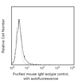
- An isotype control should be used at the same concentration as the antibody of interest.
- Caution: Sodium azide yields highly toxic hydrazoic acid under acidic conditions. Dilute azide compounds in running water before discarding to avoid accumulation of potentially explosive deposits in plumbing.
- Sodium azide is a reversible inhibitor of oxidative metabolism; therefore, antibody preparations containing this preservative agent must not be used in cell cultures nor injected into animals. Sodium azide may be removed by washing stained cells or plate-bound antibody or dialyzing soluble antibody in sodium azide-free buffer. Since endotoxin may also affect the results of functional studies, we recommend the NA/LE (No Azide/Low Endotoxin) antibody format, if available, for in vitro and in vivo use.
- Accutase is a registered trademark of Innovative Cell Technologies, Inc.
- Please refer to www.bdbiosciences.com/us/s/resources for technical protocols.
関連製品

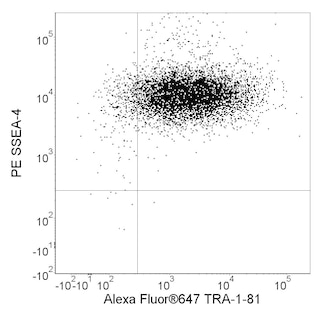
The 105/A2B5 monoclonal antibody binds to gangliosides in neural tissue in the retina, brain, spinal cord, and dorsal root ganglia. The antigenic gangliosides are found on the plasma membrane of neural cell bodies, but not axons or dendrites. Expression levels of the gangliosides that mAb 105/A2B5 binds to, including GT3 and its O-acetylated derivative, decrease during embryonic development of the brain. Thus, mAb 105/A2B5 may be used to monitor the maturation of brain tissue. A2B5 antigens have also been associated with numerous tumors, including glioblastoma multiforme tumor cells, pancreatic islet tumor cells, and rat insulinoma cells, and are thought to contribute to chemoresistance and increased tumor proliferation and recurrence.
Development References (2)
-
Balik V, Mirossay P, Bohus P, Sulla I, Mirossay L, Sarissky M. Flow cytometry analysis of neural differentiation markers expression in human glioblastomas may predict their response to chemotherapy. 2009; 29:845-858. (Clone-specific: Flow cytometry). View Reference
-
Eisenbarth GS, Walsh FS, Nirenberg M. Monoclonal antibody to a plasma membrane antigen of neurons. Proc Natl Acad Sci U S A. 1979; 76(10):4913-4917. (Immunogen: Cytotoxicity, Immunofluorescence). View Reference
Please refer to Support Documents for Quality Certificates
Global - Refer to manufacturer's instructions for use and related User Manuals and Technical data sheets before using this products as described
Comparisons, where applicable, are made against older BD Technology, manual methods or are general performance claims. Comparisons are not made against non-BD technologies, unless otherwise noted.
For Research Use Only. Not for use in diagnostic or therapeutic procedures.