Preparation and Storage
Product Notices
- This reagent has been pre-diluted for use at the recommended Volume per Test. We typically use 1 × 10^6 cells in a 100-µl experimental sample (a test).
- An isotype control should be used at the same concentration as the antibody of interest.
- Caution: Sodium azide yields highly toxic hydrazoic acid under acidic conditions. Dilute azide compounds in running water before discarding to avoid accumulation of potentially explosive deposits in plumbing.
- Source of all serum proteins is from USDA inspected abattoirs located in the United States.
- For fluorochrome spectra and suitable instrument settings, please refer to our Multicolor Flow Cytometry web page at www.bdbiosciences.com/colors.
- Please refer to www.bdbiosciences.com/us/s/resources for technical protocols.
関連製品


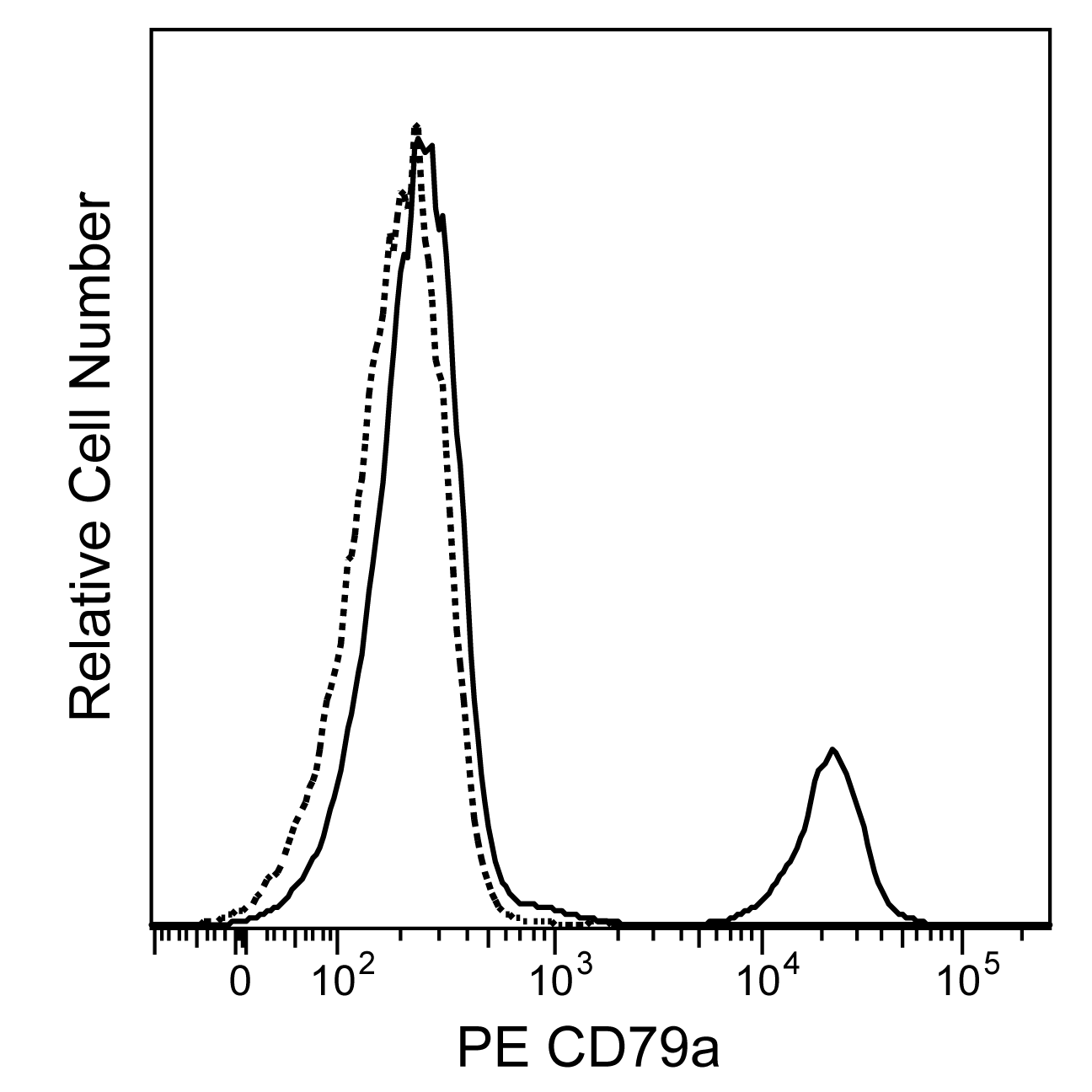
The HM57 monoclonal antibody specifically binds to the cytoplasmic domain of CD79a. CD79a is a 47-kDa type 1 transmembrane glycoprotein present on B lymphocytes. CD79a is also referred to as mb-1, IGA and Ig-alpha (Ig-α). It is expressed on B cells at various stages of differentiation, from the pre-B cell stage, probably before expression of cytoplasmic μ chain, to the plasma cell stage, in which it is detected only in the cytoplasm. CD79a associates with CD79b to form part of the B-cell receptor complex. It has been suggested that CD79a may play a role in mediating the transport of IgM to the cell surface. This antibody reportedly may crossreact with monkey, mouse, rat, bovine, canine, chicken, equine, guinea pig, porcine, and rabbit CD79a.

Development References (6)
-
Engel P, Wagner N, Tedder TF. CD79 Workshop report. In: Schlossman SF. Stuart F. Schlossman .. et al., ed. Leucocyte typing V : white cell differentiation antigens : proceedings of the fifth international workshop and conference held in Boston, USA, 3-7 November, 1993. Oxford: Oxford University Press; 1995:667-670.
-
Faldyna M, Sinkora J, Knotigova P, Rehakova Z, Moravkova A, Toman M. Flow cytometric analysis of bone marrow leukocytes in neonatal dogs. Vet Immunol Immunopathol. 2003; 95(3-4):165-176. (Clone-specific: Flow cytometry). View Reference
-
Jones M, Cordell JL, Beyers AD, Tse AG, Mason DY. Detection of T and B cells in many animal species using cross-reactive anti-peptide antibodies. J Immunol. 1993; 150(12):5429-5435. (Clone-specific: Immunohistochemistry, Western blot). View Reference
-
Mason DY, Cordell JL, Tse AG, et al. The IgM-associated protein mb-1 as a marker of normal and neoplastic B cells. J Immunol. 1991; 147(11):2474-2482. (Immunogen: Flow cytometry, Immunoaffinity chromatography, Immunofluorescence, Immunohistochemistry, Immunoprecipitation). View Reference
-
Sakaguchi N, Kashiwamura S, Kimoto M, Thalmann P, Melchers F. B lymphocyte lineage-restricted expression of mb-1, a gene with CD3-like structural properties. EMBO J. 1988; 7(11):3457-3464. (Biology). View Reference
-
Schlossman SF. Stuart F. Schlossman .. et al., ed. Leucocyte typing V : white cell differentiation antigens : proceedings of the fifth international workshop and conference held in Boston, USA, 3-7 November, 1993. Oxford: Oxford University Press; 1995.
Please refer to Support Documents for Quality Certificates
Global - Refer to manufacturer's instructions for use and related User Manuals and Technical data sheets before using this products as described
Comparisons, where applicable, are made against older BD Technology, manual methods or are general performance claims. Comparisons are not made against non-BD technologies, unless otherwise noted.
For Research Use Only. Not for use in diagnostic or therapeutic procedures.