





.png)


Preparation and Storage


推奨アッセイ手順
The PE-conjugated TN3-19.12 antibody can be used for multicolor immunofluorescent staining and flow cytometric analyses to identify and enumerate rat TNF-producing cells within mixed cell populations (see Figure). For optimal immunofluorescent staining with flow cytometric analysis, this anti-cytokine antibody should be titrated (≤ 0.5 µg mAb/million cells). For specific methodology, please visit our web site, www.bdbiosciences.com, and go to the protocols section or the chapter on intracellular staining in the Immune Function Handbook.
A useful control for demonstrating specificity of staining is either of the following: 1) pre-block the conjugated TN3-19.12 antibody with a molar excess of ligand prior to staining, or 2) pre-block the fixed/permeabilized cells with unconjugated TN3-19.12 antibody (Cat. No. 557516) prior to staining. The staining technique and blocking controls are described in detail by C. Prussin and D. Metcalfe. A suitable hamster IgG1, λ isotype control for assessing the level of background staining on paraformaldehyde-fixed/saponin-permeabilized mouse and human cells is PE-G235-2356 (Cat. No. 554711); use at comparable concentrations to antibody of interest (e.g., ≤ 0.5 µg mAb/ 1 million cells).
OTHER APPLICATIONS
In vitro neutralization: The NA/LE™ format of the TN3-19.12 antibody (Cat. No. 557370) is useful for neutralization of mouse TNF bioactivity.
IP/WB: The TN3-19.12 antibody has been reported to be useful for immunoprecipitation and Western blot. Please note that this application is not routinely tested at BD Biosciences Pharmingen.
ELISA: The purified TN3-19.12 antibody (Cat. No. 557516) can be used as a capture antibody for a sandwich ELISA that measures TNF protein levels.
Product Notices
- Since applications vary, each investigator should titrate the reagent to obtain optimal results.
- Please refer to www.bdbiosciences.com/us/s/resources for technical protocols.
- For fluorochrome spectra and suitable instrument settings, please refer to our Multicolor Flow Cytometry web page at www.bdbiosciences.com/colors.
- Caution: Sodium azide yields highly toxic hydrazoic acid under acidic conditions. Dilute azide compounds in running water before discarding to avoid accumulation of potentially explosive deposits in plumbing.
関連製品
.png?imwidth=320)

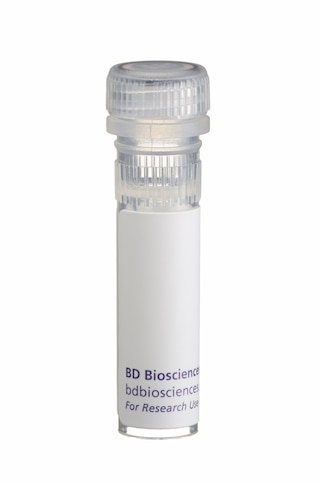
The TN3-19.12 antibody reacts with rat and mouse tumor-necrosis factor (TNF) proteins (also known as TNF-α) . Moreover, the TN3-19.12 antibody is reported to crossreact with rabbit TNF, but it does not crossreact with mouse lymphotoxin-α (LT-α, also known as TNF-β) nor with human TNF. The immunogen used to generate the TN3-19.12 hybridoma was E. coli-expressed, purified recombinant mouse TNF protein. This monoclonal antibody has been reported to neutralize the bioactivities of mouse, rat and rabbit TNF.
This antibody is routinely tested by flow cytometric analysis. Other applications were tested at BD Biosciences Pharmingen during antibody development only or reported in the literature.

Development References (7)
-
Ferran C, Dautry F, Merite S, et al. Anti-tumor necrosis factor modulates anti-CD3-triggered T cell cytokine gene expression in vivo. J Clin Invest. 1994; 93(5):2189-2196. (Clone-specific: Neutralization). View Reference
-
Leiby DA, Fortier AH, Crawford RM, Schreiber RD, Nacy CA. In vivo modulation of the murine immune response to Francisella tularensis LVS by administration of anticytokine antibodies. Infect Immun. 1992; 60(1):84-89. (Clone-specific: Neutralization). View Reference
-
Merrick BA, He CY, Craig WA, et al. Two dimensional gel electrophoresis of cellular and secreted proteins from rat alveolar macrophages after lipopolysaccharide treatment. Appl Theor Electrophor. 1992; 2(6):177-187. (Clone-specific: Immunoprecipitation, Western blot). View Reference
-
Prussin C, Metcalfe DD. Detection of intracytoplasmic cytokine using flow cytometry and directly conjugated anti-cytokine antibodies. J Immunol Methods. 1995; 188(1):117-128. (Methodology: IC/FCM Block). View Reference
-
Rabinovici R, Bugelski PJ, Esser KM, et al. Tumor necrosis factor-alpha mediates endotoxin-induced lung injury in platelet activating factor-primed rats. J Pharmacol Exp Ther. 1993; 267(3):1550-1557. (Clone-specific: Neutralization). View Reference
-
Sheehan KC, Ruddle NH, Schreiber RD. Generation and characterization of hamster monoclonal antibodies that neutralize murine tumor necrosis factors. J Immunol. 1989; 142(11):3884-3893. (Immunogen: ELISA, Immunoprecipitation, Neutralization, Western blot). View Reference
-
Takahashi S, Kapas L, Fang J, Krueger JM. An anti-tumor necrosis factor antibody suppresses sleep in rats and rabbits. Brain Res. 1995; 690(2):241-244. (Clone-specific: Neutralization). View Reference
Please refer to Support Documents for Quality Certificates
Global - Refer to manufacturer's instructions for use and related User Manuals and Technical data sheets before using this products as described
Comparisons, where applicable, are made against older BD Technology, manual methods or are general performance claims. Comparisons are not made against non-BD technologies, unless otherwise noted.
For Research Use Only. Not for use in diagnostic or therapeutic procedures.
