
Preparation and Storage
推奨アッセイ手順
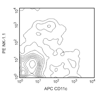
BD® CompBeads can be used as surrogates to assess fluorescence spillover (compensation). When fluorochrome conjugated antibodies are bound to BD® CompBeads, they have spectral properties very similar to cells. However, for some fluorochromes there can be small differences in spectral emissions compared to cells, resulting in spillover values that differ when compared to biological controls. It is strongly recommended that when using a reagent for the first time, users compare the spillover on cells and BD® CompBeads to ensure that BD® CompBeads are appropriate for your specific cellular application.
Product Notices
- Please refer to www.bdbiosciences.com/us/s/resources for technical protocols.
- Caution: Sodium azide yields highly toxic hydrazoic acid under acidic conditions. Dilute azide compounds in running water before discarding to avoid accumulation of potentially explosive deposits in plumbing.
- Since applications vary, each investigator should titrate the reagent to obtain optimal results.
- For fluorochrome spectra and suitable instrument settings, please refer to our Multicolor Flow Cytometry web page at www.bdbiosciences.com/colors.
- An isotype control should be used at the same concentration as the antibody of interest.
- Please observe the following precautions: We recommend that special precautions be taken (such as wrapping vials, tubes, or racks in aluminum foil) to protect exposure of conjugated reagents, including cells stained with those reagents, to any room illumination. Absorption of visible light can significantly affect the emission spectra and quantum yield of tandem fluorochrome conjugates.
- Texas Red is a registered trademark of Molecular Probes, Inc., Eugene, OR.
- CF™ is a trademark of Biotium, Inc.
- When excited by the yellow-green (561-nm) laser, the fluorescence may be brighter than when excited by the blue (488-nm) laser.
- This product is provided under an Agreement between BIOTIUM and BD Biosciences. The manufacture, use, sale, offer for sale, or import of this product is subject to one or more patents or pending applications owned or licensed by Biotium, Inc. This product, and only in the amount purchased by buyer, may be used solely for buyer’s own internal research, in a manner consistent with the accompanying product literature. No other right to use, sell or otherwise transfer (a) this product, or (b) its components is hereby granted expressly, by implication or by estoppel. This product is for research use only. Diagnostic uses require a separate license from Biotium, Inc. For information on purchasing a license to this product including for purposes other than research, contact Biotium, Inc., 3159 Corporate Place, Hayward, CA 94545, Tel: (510) 265-1027. Fax: (510) 265-1352. Email: btinfo@biotium.com.
- Because of the broad absorption spectrum of the tandem fluorochrome, extra care must be taken when using multi-laser cytometers, which may directly excite both PE and CF™594.
- Source of all serum proteins is from USDA inspected abattoirs located in the United States.
- Please refer to http://regdocs.bd.com to access safety data sheets (SDS).
- For U.S. patents that may apply, see bd.com/patents.
関連製品


.png?imwidth=320)
The URA-1 monoclonal antibody specifically recognizes the extracellular domain of the mouse CD301b. CD301b is also known as macrophage galactose-type C-type lectin 2 (MGL2) and is encoded by Mgl2 (Macrophage galactose N-acetyl-galactosamine specific lectin 2). Mouse D301b (MGL2) is a ~42 kDa type II transmembrane glycoproteins that is comprised of an extracellular region with a carbohydrate recognition domain (CRD) followed by a transmembrane region and a cytoplasmic tail. CD301b is expressed on conventional dendritic cells (cDCs) and immature DCs. CD301b recognizes molecules having galactose and N-actetylgalactosamine (GalNAc) residues. CD301b is involved in the recognition and endocytosis of glycoproteins and play roles in tissue remodeling, clearance of apoptotic cells, and defense against tumor cells. The ER-MP23 monoclonal antibody specifically recognizes both CD301a and CD301b and can reportedly inhibit their binding of carbohydrate ligands.

Development References (3)
-
Denda-Nagai K, Aida S, Saba K, et al. Distribution and function of macrophage galactose-type C-type lectin 2 (MGL2/CD301b): efficient uptake and presentation of glycosylated antigens by dendritic cells.. J Biol Chem. 2010; 285(25):19193-204. (Immunogen: Blocking, ELISA, Flow cytometry, Immunohistochemistry). View Reference
-
Sato K, Imai Y, Higashi N, et al. Lack of antigen-specific tissue remodeling in mice deficient in the macrophage galactose-type calcium-type lectin 1/CD301a.. Blood. 2005; 106(1):207-15. (Clone-specific: Immunohistochemistry). View Reference
-
Tsuiji M, Fujimori M, Ohashi Y, et al. Molecular cloning and characterization of a novel mouse macrophage C-type lectin, mMGL2, which has a distinct carbohydrate specificity from mMGL1.. J Biol Chem. 2002; 277(32):28892-901. (Biology). View Reference
Please refer to Support Documents for Quality Certificates
Global - Refer to manufacturer's instructions for use and related User Manuals and Technical data sheets before using this products as described
Comparisons, where applicable, are made against older BD Technology, manual methods or are general performance claims. Comparisons are not made against non-BD technologies, unless otherwise noted.
For Research Use Only. Not for use in diagnostic or therapeutic procedures.