







Preparation and Storage


Product Notices
- Since applications vary, each investigator should titrate the reagent to obtain optimal results.
- An isotype control should be used at the same concentration as the antibody of interest.
- Pacific Blue™ is a trademark of Molecular Probes, Inc., Eugene, OR.
- Source of all serum proteins is from USDA inspected abattoirs located in the United States.
- Caution: Sodium azide yields highly toxic hydrazoic acid under acidic conditions. Dilute azide compounds in running water before discarding to avoid accumulation of potentially explosive deposits in plumbing.
- For fluorochrome spectra and suitable instrument settings, please refer to our Multicolor Flow Cytometry web page at www.bdbiosciences.com/colors.
- Please refer to www.bdbiosciences.com/us/s/resources for technical protocols.
関連製品
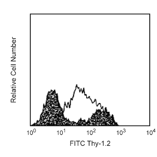




The MIH5 monoclonal antibody specifically binds to CD274, also known as B7-H1 or PDL1, a 43-kDa glycoprotein encoded by the Pdcd1lg1 gene of the B7 family of the Ig superfamily. Pdcd1lg1 mRNA is expressed in more tissues than other members of the B7 family; transcripts are found in lymphoid tissues and many, but not all, non-lymphoid tissues. The protein has been detected at low levels on resting peripheral T and B lymphocytes, macrophages, and dendritic cells. B7-H1 mRNA and protein expression are upregulated upon activation of T and B cells, macrophages, dendritic cells, and epidermal keratinocytes by a variety of stimulatory factors. B7-H1's receptor, PD-1, contains an ITIM (Immunoreceptor Tyrosine-based Inhibitory Motif) on its intracytoplasmic region and is expressed on activated B and T lymphocytes, suggesting that B7-H1-PD-1 interaction may be involved in the negative regulation of immune responses. The second PD-1 ligand, B7-DC (PD-L2), is also a member of the B7 family of the Ig superfamily. Furthermore, B7-H1 may participate in positive immunoregulation, or costimulation of T cells, through an additional receptor, which is not PD-1 and distinct from the alternate receptor for B7-DC. The MIH5 antibody blocks the binding of PD-1-Ig to B7-H1 transfectants.
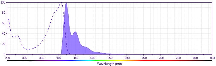
The antibody was conjugated to BD Horizon BV421 which is part of the BD Horizon Brilliant™ Violet family of dyes. With an Ex Max of 407-nm and Em Max at 421-nm, BD Horizon BV421 can be excited by the violet laser and detected in the standard Pacific Blue™ filter set (eg, 450/50-nm filter). BD Horizon BV421 conjugates are very bright, often exhibiting a 10 fold improvement in brightness compared to Pacific Blue conjugates.
Development References (9)
-
Ansari MJ, Salama AD, Chitnis T, et al. The programmed death-1 (PD-1) pathway regulates autoimmune diabetes in nonobese diabetic (NOD) mice. J Exp Med. 2003 July; 198(1):63-69. (Biology). View Reference
-
Carreno BM, Collins M. The B7 family of ligands and its receptors: New pathways for costimulation and inhibition of immune responses. Annu Rev Immunol. 2002; 20:29-53. (Biology). View Reference
-
Dong H, Chen L. B7-H1 pathway and its role in the evasion of tumor immunity. J Mol Med. 2003 May; 81(5):281-287. (Biology). View Reference
-
Hessel EM, Chu M, Lizcano JO, et al. Immunostimulatory oligonucleotides block allergic airway inflammation by inhibiting Th2 cell activation and IgE-mediated cytokine induction. J Exp Med. 2005; 202(11):1563-1573. (Clone-specific: Flow cytometry). View Reference
-
Liu X, Gao JX, Wen J, et al. B7DC/PDL2 promotes tumor immunity by a PD-1-independent mechanism. J Exp Med. 2003; 197:1721-1730. (Biology). View Reference
-
Tamura H, Dong H, Zhu G, et al. B7-H1 costimulation preferentially enhances CD28-independent T-helper cell function. Blood. 2001; 97(6):1809-1816. (Biology). View Reference
-
Tsushima F, Iwai H, Otsuki N, et al. Preferential contribution of B7-H1 to programmed death-1-mediated regulation of hapten-specific allergic inflammatory responses. Eur J Immunol. 2003; 33(10):2773-2782. (Immunogen: Blocking, Enhancement, Functional assay, Immunofluorescence, Immunohistochemistry, Inhibition, In vivo exacerbation). View Reference
-
Wang S, Bajorath J, Flies DB, Dong H, Honjo T, Chen L. Molecular modeling and functional mapping of B7-H1 and B7-DC uncouple costimulatory function from PD-1 interaction. J Exp Med. 2003; 197:1083-1091. (Biology). View Reference
-
Yamazaki T, Akiba H, Iwai H, et al. Expression of programmed death 1 ligands by murine T cells and APC. J Immunol. 2002; 169(10):5538-5545. (Biology). View Reference
Please refer to Support Documents for Quality Certificates
Global - Refer to manufacturer's instructions for use and related User Manuals and Technical data sheets before using this products as described
Comparisons, where applicable, are made against older BD Technology, manual methods or are general performance claims. Comparisons are not made against non-BD technologies, unless otherwise noted.
For Research Use Only. Not for use in diagnostic or therapeutic procedures.
