Preparation and Storage
推奨アッセイ手順
BD® CompBeads can be used as surrogates to assess fluorescence spillover (compensation). When fluorochrome conjugated antibodies are bound to BD® CompBeads, they have spectral properties very similar to cells. However, for some fluorochromes there can be small differences in spectral emissions compared to cells, resulting in spillover values that differ when compared to biological controls. It is strongly recommended that when using a reagent for the first time, users compare the spillover on cells and BD® CompBeads to ensure that BD® CompBeads are appropriate for your specific cellular application.
For optimal and reproducible results, BD Horizon Brilliant Stain Buffer should be used anytime BD Horizon Brilliant dyes are used in a multicolor flow cytometry panel. Fluorescent dye interactions may cause staining artifacts which may affect data interpretation. The BD Horizon Brilliant Stain Buffer was designed to minimize these interactions. When BD Horizon Brilliant Stain Buffer is used in in the multicolor panel, it should also be used in the corresponding compensation controls for all dyes to achieve the most accurate compensation. For the most accurate compensation, compensation controls created with either cells or beads should be exposed to BD Horizon Brilliant Stain Buffer for the same length of time as the corresponding multicolor panel. More information can be found in the Technical Data Sheet of the BD Horizon Brilliant Stain Buffer (Cat. No. 563794/566349) or the BD Horizon Brilliant Stain Buffer Plus (Cat. No. 566385).
Note: When using high concentrations of antibody, background binding of this dye to erythroid cell subsets (mature erythrocytes and precursors) has been observed. For researchers studying these cell populations, or in cases where light scatter gating does not adequately exclude these cells from the analysis, this background may be an important factor to consider when selecting reagents for panel(s).
Product Notices
- Please refer to www.bdbiosciences.com/us/s/resources for technical protocols.
- Caution: Sodium azide yields highly toxic hydrazoic acid under acidic conditions. Dilute azide compounds in running water before discarding to avoid accumulation of potentially explosive deposits in plumbing.
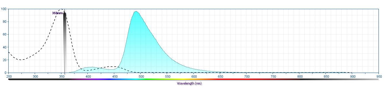
- For fluorochrome spectra and suitable instrument settings, please refer to our Multicolor Flow Cytometry web page at www.bdbiosciences.com/colors.
- BD Horizon Brilliant Ultraviolet 496 is covered by one or more of the following US patents: 8,110,673; 8,158,444; 8,227,187; 8,575,303; 8,354,239.
- BD Horizon Brilliant Stain Buffer is covered by one or more of the following US patents: 8,110,673; 8,158,444; 8,575,303; 8,354,239.
- Please refer to http://regdocs.bd.com to access safety data sheets (SDS).
- Since applications vary, each investigator should titrate the reagent to obtain optimal results.
- Human donor specific background has been observed in relation to the presence of anti-polyethylene glycol (PEG) antibodies, developed as a result of certain vaccines containing PEG, including some COVID-19 vaccines. We recommend use of BD Horizon Brilliant™ Stain Buffer in your experiments to help mitigate potential background. For more information visit https://www.bdbiosciences.com/en-us/support/product-notices.
- Researchers should determine the optimal concentration of this reagent for their individual applications.
- The production process underwent stringent testing and validation to assure that it generates a high-quality conjugate with consistent performance and specific binding activity. However, verification testing has not been performed on all conjugate lots.
関連製品


The UK98/6 monoclonal antibody specifically recognizes Urokinase-type Plasminogen Activator (uPA) that is encoded by PLAU (Plasminogen Activator Urokinase). uPA (PLAU) is secreted as an inactive single-chain precursor protein (Pro-urokinase or Pro-uPA) by various cells including vascular endothelial cells, renal epithelial cells, fibroblasts, monocytes, macrophages, smooth muscle cells and tumor cells of different origin. Following proteolytic cleavage, two resulting chains are disulfide bonded together with an amino-terminal A-chain (ATF) with EGF-like growth factor and Kringle domains bound to a catalytically active peptidase, the C-terminal B-chain. uPA (PLAU) binds to cell-surface CD87 (also known as, Urokinase Plasminogen Activator Receptor or uPA-R) by its growth factor-like domain. CD87 is widely expressed by monocytes, neutrophils, dendritic cells, activated T cells, NK cells, fibroblasts, endothelial cells, keratinocytes, and some tumor cells. The cell-associated form of uPA (PLAU) efficiently acts upon the plasminogen proenzyme to generate active plasmin which degrades blood plasma proteins and dissolves fibrin blood clots. uPA (PLAU) also activates collagenases that are involved in the breakdown of the extracellular matrix as well as some mediators of the complement system. It may play a role in tumor cell proliferation, migration, and metastases. A specific polymorphism of uPA (PLAU) is associated with late-onset Alzheimer's disease. The UK98-6 antibody can reportedly suppress the biological activity of uPA (PLAU).
Development References (6)
-
Danø K, Andreasen PA, Grøndahl-Hansen J, Kristensen P, Nielsen LS, Skriver L. Plasminogen activators, tissue degradation, and cancer.. Adv Cancer Res. 1985; 44:139-266. (Biology). View Reference
-
Finckh U, van Hadeln K, Müller-Thomsen T, et al. Association of late-onset Alzheimer disease with a genotype of PLAU, the gene encoding urokinase-type plasminogen activator on chromosome 10q22.2.. Neurogenetics. 2003; 4(4):213-7. (Biology). View Reference
-
Heidtmann HH, Hofmann M, Jacob E, Erbil C, Havemann K, Schwartz-Albiez R. Synthesis and secretion of plasminogen activators and plasminogen activator inhibitors in cell lines of different groups of human lung tumors.. Cancer Res. 1989; 49(24 Pt 1):6960-5. (Biology: Flow cytometry). View Reference
-
Meissauer A, Kramer MD, Hofmann M, et al. Urokinase-type and tissue-type plasminogen activators are essential for in vitro invasion of human melanoma cells.. Exp Cell Res. 1991; 192(2):453-9. (Clone-specific: Blocking, ELISA, Functional assay). View Reference
-
Meissauer A, Kramer MD, Schirrmacher V, Brunner G. Generation of cell surface-bound plasmin by cell-associated urokinase-type or secreted tissue-type plasminogen activator: a key event in melanoma cell invasiveness in vitro.. Exp Cell Res. 1992; 199(2):179-90. (Clone-specific: Blocking, ELISA, Functional assay). View Reference
-
Schwartz-Albiez R, Heidtmann HH, Wolf D, Schirrmacher V, Moldenhauer G. Three types of human lung tumour cell lines can be distinguished according to surface expression of endogenous urokinase and their capacity to bind exogenous urokinase.. Br J Cancer. 1992; 65(1):51-7. (Immunogen: ELISA, Radioimmunoassay, Western blot). View Reference
Please refer to Support Documents for Quality Certificates
Global - Refer to manufacturer's instructions for use and related User Manuals and Technical data sheets before using this products as described
Comparisons, where applicable, are made against older BD Technology, manual methods or are general performance claims. Comparisons are not made against non-BD technologies, unless otherwise noted.
For Research Use Only. Not for use in diagnostic or therapeutic procedures.