





.png)


Preparation and Storage


Product Notices
- Since applications vary, each investigator should titrate the reagent to obtain optimal results.
- An isotype control should be used at the same concentration as the antibody of interest.
- Please refer to www.bdbiosciences.com/us/s/resources for technical protocols.
- Caution: Sodium azide yields highly toxic hydrazoic acid under acidic conditions. Dilute azide compounds in running water before discarding to avoid accumulation of potentially explosive deposits in plumbing.
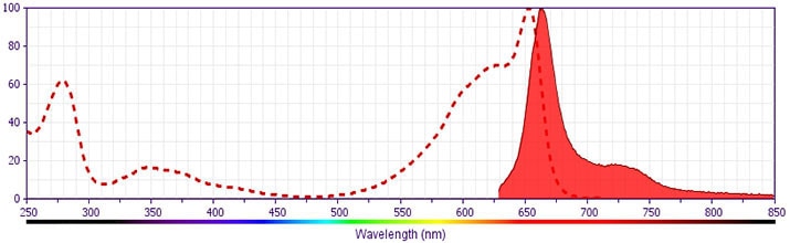
- This APC-conjugated reagent can be used in any flow cytometer equipped with a dye, HeNe, or red diode laser.
- For fluorochrome spectra and suitable instrument settings, please refer to our Multicolor Flow Cytometry web page at www.bdbiosciences.com/colors.
関連製品

.png?imwidth=320)
The APA5 antibody monoclonal antibody specifically binds to CD140a, the Platelet-Derived Growth Factor Receptor alpha chain (PDGFR-a, PDGFR-α). CD140a is a receptor tyrosine kinase that is widely expressed on cells of mesenchymal origin in the embryo and adult. CD140a is expressed on several other cell types during embryonic development, but not on hematopoietic cells. CD140a binds to PDGF A and B chains, in contrast to PDGFR-b (CD140b) that binds only to the PDGF B chain. Biologically active PDGF is a disulphide-linked dimer, forming the AA, AB, and BB isoforms. Ligand binding to the PDGF Receptor induces the formation of receptor dimers (aa, ab, or bb), autophosphorylation, and internalization. The APA5 antibody has been demonstrated to block binding of PDGF-AA to PDGFR-a-expressing cells in vitro and to block some PDGF-mediated developmental events in vivo.
Development References (3)
-
Fruttiger M, Calver AR, Krüger WH. PDGF mediates a neuron-astrocyte interaction in the developing retina. Neuron. 1996; 17(6):1117-1131. (Clone-specific: Blocking). View Reference
-
Heldin CH. Structural and functional studies on platelet-derived growth factor. EMBO J. 1992; 11(12):4251-4259. (Biology). View Reference
-
Takakura N, Yoshida H, Kunisada T, Nishikawa S, Nishikawa SI. Involvement of platelet-derived growth factor receptor-alpha in hair canal formation. J Invest Dermatol. 1996; 107(5):770-777. (Immunogen: Blocking, ELISA, Immunohistochemistry, Inhibition, Western blot). View Reference
Please refer to Support Documents for Quality Certificates
Global - Refer to manufacturer's instructions for use and related User Manuals and Technical data sheets before using this products as described
Comparisons, where applicable, are made against older BD Technology, manual methods or are general performance claims. Comparisons are not made against non-BD technologies, unless otherwise noted.
For Research Use Only. Not for use in diagnostic or therapeutic procedures.
