BD Pharmingen™ Alexa Fluor® 647 Mouse Anti-Human HLA-DR, DP, DQ
クローン Tu39 (also known as TÜ39)
(RUO)

Preparation and Storage
Product Notices
- This reagent has been pre-diluted for use at the recommended Volume per Test. We typically use 1 × 10^6 cells in a 100-µl experimental sample (a test).
- Source of all serum proteins is from USDA inspected abattoirs located in the United States.
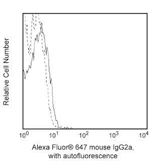
- An isotype control should be used at the same concentration as the antibody of interest.
- The Alexa Fluor®, Pacific Blue™, and Cascade Blue® dye antibody conjugates in this product are sold under license from Molecular Probes, Inc. for research use only, excluding use in combination with microarrays, or as analyte specific reagents. The Alexa Fluor® dyes (except for Alexa Fluor® 430), Pacific Blue™ dye, and Cascade Blue® dye are covered by pending and issued patents.
- Alexa Fluor® is a registered trademark of Molecular Probes, Inc., Eugene, OR.
- Alexa Fluor® 647 fluorochrome emission is collected at the same instrument settings as for allophycocyanin (APC).
- Caution: Sodium azide yields highly toxic hydrazoic acid under acidic conditions. Dilute azide compounds in running water before discarding to avoid accumulation of potentially explosive deposits in plumbing.
- For fluorochrome spectra and suitable instrument settings, please refer to our Multicolor Flow Cytometry web page at www.bdbiosciences.com/colors.
- Species cross-reactivity detected in product development may not have been confirmed on every format and/or application.
- Please refer to www.bdbiosciences.com/us/s/resources for technical protocols.
関連製品


The TU39 monoclonal antibody specifically recognizes human major histocompatibility (MHC) Class II HLA-DR, DP and most DQ antigens. These antigens are encoded by genes within the Human Leukocyte Antigen (HLA) Complex located on chromosome 6. MHC Class II antigens are transmembrane heterodimeric glycoproteins composed of α chain (36 kDa) and β chain (27 kDa) subunits. They are expressed primarily on antigen presenting cells which include dendritic cells, monocytes, macrophages, thymic epithelial cells, and B cells. They are also expressed on activated T cells. This molecule plays a major role in mediating cellular interactions during antigen presentation to CD4+ T lineage cells. The TU39 antibody is reportedly useful for immunophenotyping as well as functional studies including the inhibition of mixed lymphocyte reactions and antibody-mediated complement fixation on target cells.
Development References (4)
-
Barclay NA, Brown MH, Birkeland ML, et al, ed. The Leukocyte Antigen FactsBook. San Diego, CA: Academic Press; 1997.
-
Pawelec G, Ziegler A, Wernet P. Dissection of human allostimulatory determinants with cloned T cells: stimulation inhibition by monoclonal antibodies TU22, 34, 35, 36, 37, 39, 43, and 58 against distinct human MHC class II molecules. Hum Immunol. 1985; 12(3):165-176. (Clone-specific). View Reference
-
Pawelec GP, Shaw S, Ziegler A, Muller C, Wernet P. Differential inhibition of HLA-D- or SB-directed secondary lymphoproliferative responses with monoclonal antibodies detecting human Ia-like determinants. J Immunol. 1982; 129(3):1070-1075. (Clone-specific: Cytotoxicity, Immunoprecipitation, Inhibition). View Reference
-
Ziegler A, Heinig J, Muller C, et al. Analysis by sequential immunoprecipitations of the specificities of the monoclonal antibodies TU22,34,35,36,37,39,43,58 and YD1/63.HLK directed against human HLA class II antigens. Immunobiology. 1986; 171(1-2):77-92. (Clone-specific). View Reference
Please refer to Support Documents for Quality Certificates
Global - Refer to manufacturer's instructions for use and related User Manuals and Technical data sheets before using this products as described
Comparisons, where applicable, are made against older BD Technology, manual methods or are general performance claims. Comparisons are not made against non-BD technologies, unless otherwise noted.
For Research Use Only. Not for use in diagnostic or therapeutic procedures.