BD Pharmingen™ Purified Mouse Anti-Human Perlecan
克隆 5D7-2E4 (RUO)
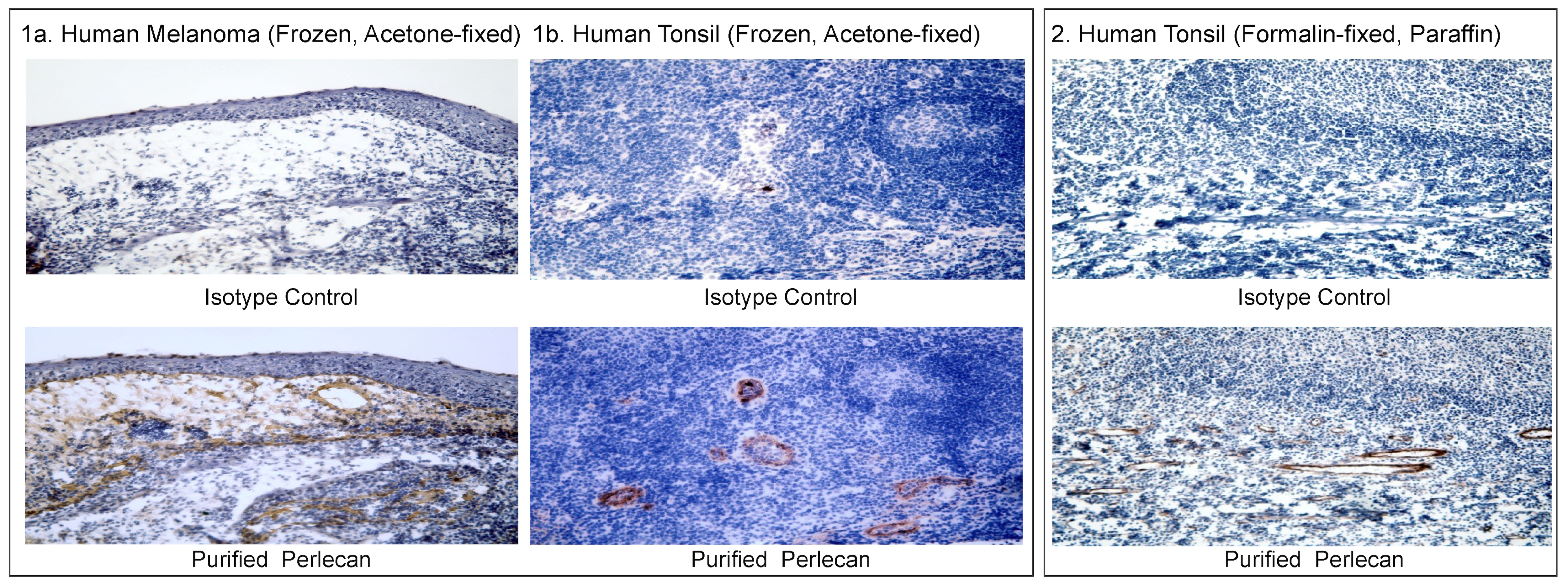
Immunohistochemical analysis of Perlecan expression by cells within human melanoma or tonsil sections.
Panel 1 - Frozen, acetone-fixed human melanoma (Panel 1a.) or tonsil (Panel 1b.) sections were stained with either Purified Mouse IgG1, κ Isotype Control (Cat.No.550878; Top Images) or Purified Mouse Anti-Human Perlecan antibody (Cat. No. 565781; Bottom Images). A three-step staining procedure that employed Biotin Goat Anti-Mouse Immunoglobulin (Cat. No. 550337), Streptavidin-Horseradish Peroxidase (HRP) (Cat. No. 550946), and the DAB Substrate Kit (Cat. No. 550880) was used to develop the primary staining reagents. Basement membranes showed positive staining. Original magnification, 20×.
Panel 2 - Following antigen retrieval with BD Retrievagen A Buffer (Cat. No. 550524) and using other IHC reagents the same as above, formalin-fixed, paraffin-embedded human tonsil sections were stained overnight with either Purified Mouse IgG1, κ Isotype Control (Top Image) or Purified Mouse Anti-Human Perlecan antibody (Bottom Image). A three-step staining procedure, that employed Biotin Goat Anti-Mouse Immunoglobulin, Streptavidin-Horseradish Peroxidase (HRP), and the DAB Substrate Kit, was used to develop the primary staining reagents. The basement membrane showed positive staining. Original magnification, 20×.
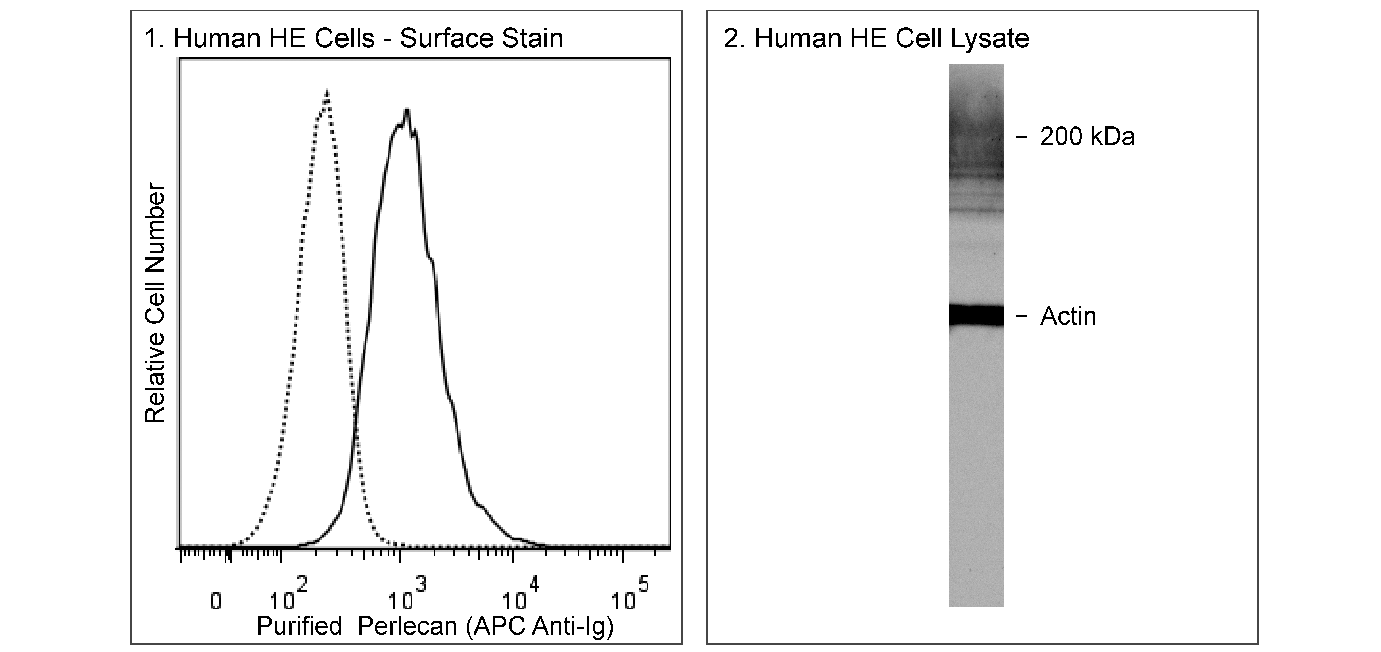
Flow cytometric and Western blot analyses of human Perlecan expression.
Panel 1 - Human HE cells were stained with either Purified Mouse IgG1, κ Isotype Control (Cat.No.554121; dashed line histogram) or Purified Mouse Anti-Human Perlecan antibody (Cat. No. 565781; solid line histogram).The cells were washed and then secondarily stained with APC Goat Anti-Mouse Ig (Cat. No. 550826). The fluorescence histogram showing surface Perlecan expression (or Ig Isotype control staining) was derived from gated events with the forward and side light-scatter characteristics of viable cells. Flow cytometric analysis was performed using a BD FACSCanto™ II Flow Cytometer System.
Panel 2 - Lysate prepared from HE cells was electrophoresed (SDS-PAGE), transferred to a PVDF membrane, and probed with Purified Anti-Human Perlecan antibody (10 μg/ml) along with Purified Mouse Anti-Actin Ab-5 (Cat. No. 612656/612657) as a control. Perlecan was detected after subsequent labeling with HRP Goat Anti-Rat Ig (Cat. No. 554017), and detection with ECL Western blotting detection reagents. Perlecan was identified in protein bands of high molecular weight.

Immunohistochemical analysis of Perlecan expression by cells within human melanoma or tonsil sections.
Panel 1 - Frozen, acetone-fixed human melanoma (Panel 1a.) or tonsil (Panel 1b.) sections were stained with either Purified Mouse IgG1, κ Isotype Control (Cat.No.550878; Top Images) or Purified Mouse Anti-Human Perlecan antibody (Cat. No. 565781; Bottom Images). A three-step staining procedure that employed Biotin Goat Anti-Mouse Immunoglobulin (Cat. No. 550337), Streptavidin-Horseradish Peroxidase (HRP) (Cat. No. 550946), and the DAB Substrate Kit (Cat. No. 550880) was used to develop the primary staining reagents. Basement membranes showed positive staining. Original magnification, 20×.
Panel 2 - Following antigen retrieval with BD Retrievagen A Buffer (Cat. No. 550524) and using other IHC reagents the same as above, formalin-fixed, paraffin-embedded human tonsil sections were stained overnight with either Purified Mouse IgG1, κ Isotype Control (Top Image) or Purified Mouse Anti-Human Perlecan antibody (Bottom Image). A three-step staining procedure, that employed Biotin Goat Anti-Mouse Immunoglobulin, Streptavidin-Horseradish Peroxidase (HRP), and the DAB Substrate Kit, was used to develop the primary staining reagents. The basement membrane showed positive staining. Original magnification, 20×.
Flow cytometric and Western blot analyses of human Perlecan expression.
Panel 1 - Human HE cells were stained with either Purified Mouse IgG1, κ Isotype Control (Cat.No.554121; dashed line histogram) or Purified Mouse Anti-Human Perlecan antibody (Cat. No. 565781; solid line histogram).The cells were washed and then secondarily stained with APC Goat Anti-Mouse Ig (Cat. No. 550826). The fluorescence histogram showing surface Perlecan expression (or Ig Isotype control staining) was derived from gated events with the forward and side light-scatter characteristics of viable cells. Flow cytometric analysis was performed using a BD FACSCanto™ II Flow Cytometer System.
Panel 2 - Lysate prepared from HE cells was electrophoresed (SDS-PAGE), transferred to a PVDF membrane, and probed with Purified Anti-Human Perlecan antibody (10 μg/ml) along with Purified Mouse Anti-Actin Ab-5 (Cat. No. 612656/612657) as a control. Perlecan was detected after subsequent labeling with HRP Goat Anti-Rat Ig (Cat. No. 554017), and detection with ECL Western blotting detection reagents. Perlecan was identified in protein bands of high molecular weight.
准备和存储
商品通知
- Since applications vary, each investigator should titrate the reagent to obtain optimal results.
- Please refer to www.bdbiosciences.com/us/s/resources for technical protocols.
- Sodium azide is a reversible inhibitor of oxidative metabolism; therefore, antibody preparations containing this preservative agent must not be used in cell cultures nor injected into animals. Sodium azide may be removed by washing stained cells or plate-bound antibody or dialyzing soluble antibody in sodium azide-free buffer. Since endotoxin may also affect the results of functional studies, we recommend the NA/LE (No Azide/Low Endotoxin) antibody format, if available, for in vitro and in vivo use.
- Caution: Sodium azide yields highly toxic hydrazoic acid under acidic conditions. Dilute azide compounds in running water before discarding to avoid accumulation of potentially explosive deposits in plumbing.
- An isotype control should be used at the same concentration as the antibody of interest.
配套商品


The 5D7-2E4 monoclonal antibody specifically recognizes the protein core of Perlecan (PRCAN), which has a calculated molecular mass of ~460 kDa. Smaller molecular mass species of Perlecan have also been described. Perlecan is also known as Heparan sulfate proteoglycan 2 (HSPG2), or Basement membrane-specific heparan sulfate proteoglycan core protein (HSPG). Perlecan is a large multidomain proteoglycan that is produced by epithelial and endothelial cells. It is a major component of the vascular extracellular matrix and helps sustain endothelial barrier function. Perlecan inhibits smooth muscle cell proliferation and serves as an attachment substrate for cells. It plays essential roles in normal cartilage, heart and vascular development, as well as, in the response to vascular injury.
研发参考 (5)
-
Chuang CY, Degendorfer G, Hammer A, Whitelock JM, Malle E, Davies MJ. Oxidation modifies the structure and function of the extracellular matrix generated by human coronary artery endothelial cells.. Biochem J. 2014; 459(2):313-22. (Immunogen: ELISA). 查看参考
-
Jung M, Lord MS, Cheng B, et al. Mast cells produce novel shorter forms of perlecan that contain functional endorepellin: a role in angiogenesis and wound healing.. J Biol Chem. 2013; 288(5):3289-304. (Clone-specific: Functional assay, Inhibition). 查看参考
-
Kallunki P, Tryggvason K.. Human basement membrane heparan sulfate proteoglycan core protein: a 467-kD protein containing multiple domains resembling elements of the low density lipoprotein receptor, laminin, neural cell adhesion molecules, and epidermal growth factor.. J Cell Biol. 1992; 116(2):559-571. (Biology). 查看参考
-
Lord MS, Chuang CY, Melrose J, Davies MJ, Iozzo RV, Whitelock JM. The role of vascular-derived perlecan in modulating cell adhesion, proliferation and growth factor signaling.. Matrix Biol. 2014; 35:112-22. (Clone-specific: ELISA). 查看参考
-
Murdoch AD, Dodge GR, Cohen I, Tuan RS, Iozzo RV.. Primary structure of the human heparan sulfate proteoglycan from basement membrane (HSPG2/perlecan). A chimeric molecule with multiple domains homologous to the low density lipoprotein receptor, laminin, neural cell adhesion molecules, and epidermal growth factor.. J Biol Chem. 1992; 267(12):8544-8557. (Biology). 查看参考
Please refer to Support Documents for Quality Certificates
Global - Refer to manufacturer's instructions for use and related User Manuals and Technical data sheets before using this products as described
Comparisons, where applicable, are made against older BD Technology, manual methods or are general performance claims. Comparisons are not made against non-BD technologies, unless otherwise noted.
For Research Use Only. Not for use in diagnostic or therapeutic procedures.