多种刺激(如细胞因子治疗),可以影响细胞增殖。细胞增殖可以在多种刺激下发生,比如细胞因子暴露或各种其他进程。
细胞增殖染料
BD生物科学公司提供了BD Horizon™ 紫色增殖染料450(VPD)和BD Horizon™ CFSE,分别采用紫色激光和蓝色激光来检测细胞增殖,这便于使用更大的检测组套(panel)。这使得通过多色流式细胞仪从有限的样本中测定更多的数据,成为可能。
这两种增殖染料,都是非荧光酯化染料。酯基允许染料进入细胞。一旦染料进入细胞内,酯酶将把酯基裂解,将染料转化为荧光产物,并将其困在细胞内。随着每次复制事件的发生,细胞中的染料量减少,导致生成一种特征图。

BD Horizon™细胞增殖染料自由进入一个细胞。一旦进入细胞,染料被非特异性酯酶裂解,释放出荧光分子,并且被困在细胞内。


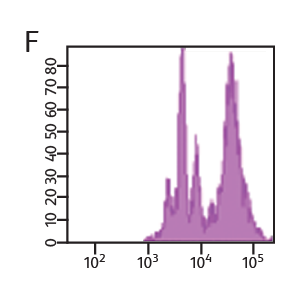
Concentration of VPD450 and cell-cycle kinetics on mouse spleen stimulated with anti-CD3e and anti-CD28. C57 Black/6 splenocytes were either loaded with varying concentrations of BD Horizon™ VPD450, DMSO, or left as untreated controls, then stimulated with anti-CD3e and anti-CD28 for two days. Cells were pulsed with BrdU prior to harvesting, then stained with APC anti-BrdU and 7-AAD (Cat. No. 552598). The top panels (A–C) illustrate APC antiBrdU and 7-AAD staining. The bottom panels (D–F) illustrate the corresponding VPD450 histograms. The control cells (Cells– ) (Panel A) and the 1-μM VPD450- loaded cell population (Panel C) demonstrated a similar percentage of BrdU+ cells (40.7% and 39.8%, respectively). Higher concentrations of dye can negatively impact cell proliferation (data not shown). To confirm that the DMSO (which is used as a solvent for VPD450) is not responsible for a decrease in proliferation, a DMSO group was included (Panel B). DMSO-treated cells incorporated a similar percentage of BrdU compared to the Cells– group and the 1-μM VPD450-loaded cell populations.
Measurement of cell proliferation with BrdU
BD Biosciences carries a series of antibodies and kits designed for the detection of proliferating cells by measurement of bromodeoxyuridine (BrdU), an analog of the DNA precursor thymidine used to measure de novo DNA synthesis.
During the S phase of the cell cycle (DNA synthesis), BrdU is incorporated into the newly synthesized DNA and can be readily detected by anti-BrdU specific antibodies. BD antibodies and kits designed for the detection of BrdU are available for both intracellular flow cytometry and immunohistochemistry and include BD Horizon™ V450, BD Horizon Brilliant Violet™ 510 (BV510), PerCP-Cy5.5 and other formats.
In addition to DNA increases, levels of certain proteins also rise as a result of cell proliferation. For example, Ki67 is an antigen that is expressed in the nucleus of dividing cells. However, during the G0 phase of the cell cycle, it is not detected. Ki67 can be combined with other proliferation markers such as BrdU and VPD450 for added confidence. These markers can also be combined with cell surface and other types of markers to gain additional information about cell subsets and their signaling pathways.
Cell proliferation analysis of mouse splenocytes


CD4+ enriched mouse splenocytes were loaded with 1 µM VPD450 for 10 minutes. Cells were then stimulated with anti-CD3/CD28 and harvested at the indicated times. Approximately 4 to 6 hours prior to harvest, cells were stimulated with PMA/ionomycin in the presence of BD GolgiStop™ Protein Transport Inhibitor. Cells were fixed and permeabilized, stained for IL-2, and analyzed on a BD® LSR II Flow Cytometer.
-
Application Notes

-
Brochure
-
Product Information Sheets
-
Webinars
采用抗CD3e和抗CD28进行刺激的小鼠脾上VPD450浓度及细胞周期动力学。C57 Black/6脾细胞,要么加载不同浓度的BD Horizon™VPD450、DMSO,要么作为未处理的对照物,然后用抗CD3e和抗CD28刺激两天。在收集之前,细胞受到BrdU的脉冲作用,然后采用APC抗BrdU和7-AAD(Cat. No. 552598)进行染色。顶部检测组套(panel)(A-C)显示了APC 抗BrdU和7-AAD的染色。底部检测组套(panel)(D-F)显示了对应的VPD450直方图。对照细胞(Cells-)(检测组套A)和1-μM VPD450-加载细胞群(检测组套C)显示 BrdU+细胞的比例相似,(分别为40.7%和39.8%)。高浓度的染料,会对细胞增殖产生负面影响(数据未显示)。为了确认DMSO(用作VPD450的溶剂)不会导致增殖减少,我们添加了DMSO组(检测组套B)。经DMSO处理的细胞,掺入了BrdU,掺入的比例,与Cell-组和1-μM VPD450-加载细胞群的BrdU比例相似。
采用BrdU测定细胞增殖
BD生物科学公司提供了一系列抗体和试剂盒,用于通过测定溴脱氧尿苷(BrdU)来检测增殖细胞,而BrdU是一种DNA前体胸腺嘧啶的类似物,用于检测新生的DNA合成。
在细胞周期的S阶段(DNA合成),BrdU被掺入到新合成的DNA中,从而可以很容易被抗BrdU特异性抗体检测到。设计用于BrdU检测的BD抗体和试剂盒,在细胞内流式细胞术和免疫组织化学染色法中均可以使用,并且包括BD Horizon™ V450、BD Horizon Brilliant Violet™ 510(BV510)、 PerCP-Cy5.5等格式。
除了DNA增加之外,某些蛋白质的水平也会随着细胞增殖而上升。例如,Ki67是一种在分裂细胞细胞核中表达的抗原。然而,在细胞周期的G0阶段,没有检测到它。Ki67可以与其他增殖标志物(如BrdU和VPD450)结合使用,以增加可信度。这些标记物还可以与细胞表面和其他类型的标记物结合,以获得关于细胞亚群及其信号传导途径方面的额外信息。
小鼠脾细胞的细胞增殖分析
将1µM VPD450加载于CD4+富集的小鼠脾细胞10分钟。然后用抗CD3/CD28刺激细胞,并且在指定时间收集细胞。在收获前约4 - 6小时,在BD GolgiStop™蛋白转运抑制剂存在的情况下,使用PMA/离子霉素刺激细胞。对细胞进行固定和破膜,然后采用IL-2染色,并且在BD® LSR II流式细胞仪上进行分析。
仅供研究使用,不用于诊疗程序。
Cy是全球生命科学解决方案德国股份有限公司的商标,或者以Cytiva名义开展业务的附属公司。