
Intracellular flow cytometry is a powerful technique for the identification of cell types and the analysis of signaling and functional responses within cell lines and heterogeneous cell samples. For some cell types, such as Th17 and regulatory T cells (Tregs), definitive identification depends on the combined use of surface and intracellular markers such as cytokines or transcription factors. Intracellular flow cytometry also provides rich information concerning cellular function and signaling responses. Fluorescent antibodies specific for cell surface markers can be combined with markers of apoptosis, proliferation and protein phosphorylation to determine which cell subsets respond to various stimuli or treatments. The combined use of multiple markers decreases data acquisition time and conserves precious samples, since more parameters can be measured on a per-cell basis. While western blotting and other methods are useful for the examination of single proteins expressed by entire cell populations, flow cytometry allows the detection of multiple proteins simultaneously at the level of individual cells.
BD Biosciences provides fluorochrome-conjugated antibodies, buffers, kits and protocols to facilitate intracellular flow cytometry. Our antibodies are tested in biologically relevant model systems. These established tools enable new discoveries in fields such as immunology, inflammation and stem cell biology. Find the tools and techniques, including BD fluorochrome-conjugated antibodies, buffers, kits and protocols that support intracellular cytokine staining and phosphoprotein and transcription factor detection by intracellular flow cytometry.
Protocols for intracellular flow cytometry
细胞内流式细胞术是鉴定细胞类型和分析细胞系和异质性细胞样本内信号传导和功能反应的有力技术。对于一些细胞类型,如Th17和调节性T细胞(Treg),其最终鉴定依赖于细胞表面和细胞内标记物的组合使用,如细胞因子或转录因子。细胞内流式细胞术还可提供细胞功能和信号反应方面的丰富信息。细胞表面标记物的特异性荧光抗体可与凋亡、增殖和蛋白磷酸化标记物结合,以确定哪些细胞亚群对各种刺激或治疗有反应。多个标记物的联合使用可减少数据采集时间并保存宝贵样本,因为这样可以测定单个细胞的更多参数。虽然蛋白免疫印迹法和其他方法都可以检测整个细胞群表达的单个蛋白,但流式细胞术可在单个细胞水平同时检测多个蛋白。
BD生物科学公司提供荧光染料标记抗体、缓冲液、试剂盒和实验方案,以促进细胞内流式细胞术的实施。已在生物学相关模型系统测试了我们的抗体。这些已确定的工具能够让我们在免疫学、炎症和干细胞生物学等领域有新的发现。找到支持通过细胞内流式细胞术检测细胞内细胞因子染色以及磷蛋白质和转录因子的工具和技术,包括BD荧光染料标记抗体、缓冲液、试剂盒和实验方案。
细胞内染色的基本原理
虽然细胞表面染色技术是相对标准的,但是,细胞内标记物的最佳染色往往取决于目标蛋白的生物学特性。根据蛋白质在细胞内的位置、与其他分子的结合及其稳定性,推荐不同的细胞制备和染色方法。例如,细胞因子是典型的分泌蛋白。
但是,如果捕获在细胞内,可以使用蛋白转运抑制剂,如BD GolgiStop™(含莫能菌素)或BD GolgiPlug™(含布雷菲德菌素A),将它们染色为细胞内蛋白。使用BD Cytofix/ Cytoperm™固定和透化溶液进行温和固定和透化后可以相对容易地获得细胞因子。
相较于细胞因子,转录因子通常位于细胞核内,并与DNA和其他蛋白质结合。一些蛋白的磷酸化(如信号转导及转录激活因子5)会导致二聚体形成,这会掩盖相关的磷酸化表位。此外,细胞内的磷酸酶可以迅速使这些蛋白质去磷酸化。因此,处理后必须迅速固定细胞并进行更强的透化,以使抗体进入细胞核,并达到被破坏的分子复合物内的表位。
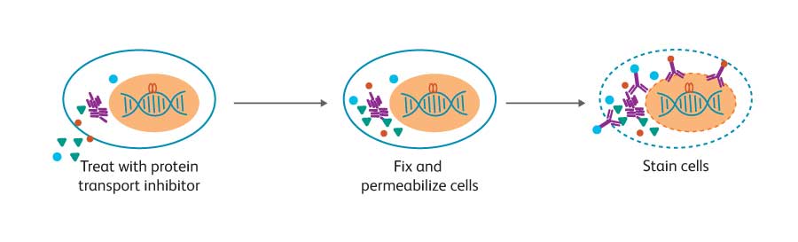
固定并透化细胞(用虚线膜表示),然后染色,并采用流式细胞术进行分析。为了研究细胞因子的分泌,首先用蛋白质运输抑制剂处理细胞,从而使靶蛋白在细胞内蓄积。
While techniques for cell surface staining are relatively standard, optimal staining for intracellular markers often depends on the biology of the target protein. Depending on the protein’s location inside the cell, association with other molecules, and its stability, different cell preparation and staining methods are recommended. Cytokines, for example, are typically secreted proteins.
However, if they are trapped inside the cell, they can be stained as intracellular proteins using protein transport inhibitors such as BD GolgiStop™ (containing monensin) or BD GolgiPlug™ (containing brefeldin A). Cytokines are relatively accessible using the gentle fixation and permeabilization afforded by BD Cytofix/ Cytoperm™ Fixation and Permeabilization Solution.
In contrast to cytokines, transcription factors often are localized inside the nucleus and bound to DNA and other proteins. Phosphorylation of some proteins, such as Stat5, results in dimer formation that masks the phosphorylated epitope of interest. Also, intracellular phosphatases can quickly dephosphorylate these proteins. Therefore, after treatment, cells must be quickly fixed and subjected to stronger permeabilization conditions to allow the antibody to enter the nucleus and access the epitope within disrupted molecular complexes.
Cells are fixed and permeabilized (symbolized by dashed line membrane), stained, and analyzed by flow cytometry. For studies on cytokine production, cells are first treated with a protein transport inhibitor to allow accumulation of the target protein inside the cell.
BD Biosciences provides tools to support intracellular flow cytometry
To facilitate intracellular flow cytometry assays, BD has developed several kits, buffers and protocols. In addition, many fluorescent antibodies specific for key cell surface markers have been tested in several buffer systems to save time, sample and money. Buffers are available for:
Detection of cytokines
BD Cytofix/Cytoperm™ Fixation/Permeabilization Solution (Cat. No. 554714) is suitable for staining most cytokines and cell surface markers. This buffer system can also be used to stain some transcription factors and other intracellular proteins. This buffer system contains mild detergents along with a formaldehyde-based fixative.
Transcription factors
The BD Pharmingen™ Transcription Factor Buffer Set (Cat. No. 562574/562725) is designed for the staining of transcription factors alone or in combination with cell surface markers and cytokines. This buffer system contains mild detergents along with a formaldehyde-based fixative.
Detection of phosphorylated protein
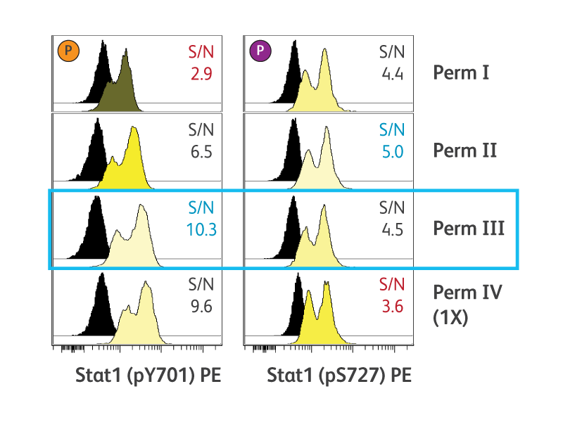
BD Phosflow™ Perm Buffer III (Cat. No. 558050) is the recommended permeabilization buffer for phosphoepitope detection by flow cytometry. Perm buffer III is a harsh alcohol-based buffer. Alternative permeabilization buffers also are available to accommodate particular experimental requirements. We also provide a convenient buffer compatibility tool, detailing many common clones and their compatibility with BD Phosflow™ Buffer protocols.
The permeabilization technique used can negatively impact the detection of cell surface and other intracellular antigens. The same techniques that allow access to the nucleus and open up DNA/protein or protein/protein complexes can often denature cell surface antigens, preventing their detection by antibodies. While detection of different intracellular proteins might require different conditions, the basic principles are the same: cells are fixed and permeabilized and then stained intracellularly with fluorescent antibodies.
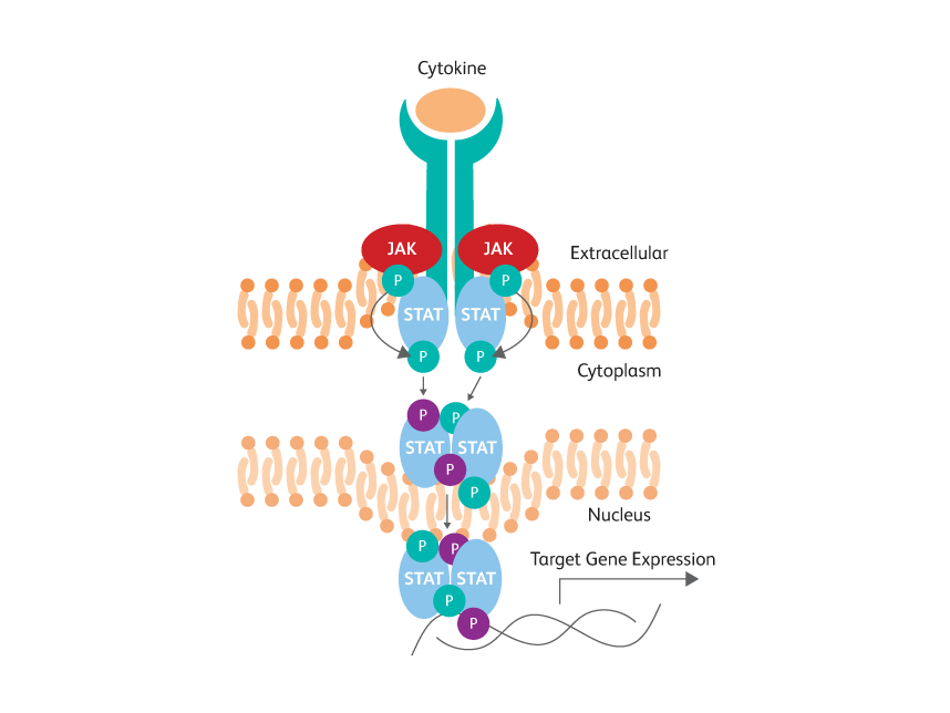
STAT phosphotyrosine epitopes are obscured by dimerization within activated cells.
Optimal cell permeabilization conditions vary by epitope location.
BD生物科学公司提供了实施细胞内流式细胞术的工具。
为了便于完成细胞内流式细胞术检测,BD公司还开发了几种试剂盒、缓冲液和实验方案。此外,已经在几个缓冲系统中对许多关键细胞表面标记物的特异性荧光抗体中进行测试,以节省时间、样品和资金。缓冲液可用于:
细胞因子的检测
BD Cytofix/Cytoperm™固定/透化液(目录号:554722)适用于染色大多数细胞因子和细胞表面标记物。该缓冲系统也可用于染色一些转录因子和其他细胞内蛋白。该缓冲系统含有温和的洗涤剂与甲醛基固定剂。
转录因子
BD Pharmingen™转录因子缓冲液(目录号:562574/562725)用于单独或联合细胞表面标记物和细胞因子对转录因子进行染色。该缓冲系统含有温和的洗涤剂与甲醛基固定剂。
磷酸化蛋白的检测
BD Phosflow™透化缓冲液III(目录号:558050)是推荐用于流式细胞术检测磷表位的透化缓冲液。透化缓冲液III是一种粗糙醇基缓冲液。对于特定的实验要求,也可使用其他符合要求的透化缓冲液。我们还提供了方便的缓冲液兼容工具,其中详细介绍了许多常见克隆及其与BD Phosflow™缓冲液实验方案的兼容性。
使用的透化技术会对细胞表面和其他细胞内抗原的检测产生负面影响。可以进入细胞核并打开DNA/蛋白质或蛋白质/蛋白质复合物的相同技术通常可以使细胞表面抗原变性,防止被抗体检测到。虽然检测不同的细胞内蛋白可能需要不同的条件,但基本原理是相同的,即固定并透化细胞,然后用荧光抗体在细胞内染色。
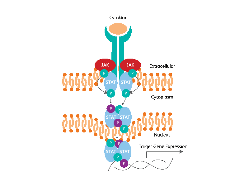
活化细胞内的STAT磷酸络氨酸表位会被二聚作用掩盖。
最佳胞透化条件因表位位置而异。
细胞因子检测
激活的各种细胞类型可以分泌细胞因子、趋化因子和其他炎症介质,如穿孔素和颗粒酶,作为免疫或炎症反应的一部分。
ELISA和BD®Cytometric Bead Array(CBA)等方法可测定全细胞群产生的分泌蛋白。相比而言,细胞内流式细胞术可分析相关细胞群内单一表型鉴定细胞类型分泌的细胞因子和其他炎症介质。
通过细胞内流式细胞术可以很容易地确定活化细胞群分泌的细胞因子是少数细胞分泌大量细胞因子的结果,还是大量细胞群每个细胞分泌少量细胞因子的结果。此外,通过细胞内流式细胞术可便于同时测量多种细胞因子,以识别多功能细胞。1细胞内细胞因子染色对多种研究都有用,包括B细胞和T细胞的分化和可塑性。
由于细胞因子通常是分泌蛋白质,必须首先使用蛋白质运输抑制剂将其捕获在细胞内。两种最常用的蛋白转运抑制剂为莫能菌素(BD GolgiStop™抑制剂)和布雷菲德菌素A(BD GolgiPlug™抑制剂)。莫能菌素可与高尔基体跨膜Na++/H+转运相互作用阻止蛋白质的分泌,而布雷菲德菌素A将细胞内分泌的蛋白质从cis/高尔基体中间复合物重新分配到内质网。2因此,蛋白质转运抑制剂的最佳选择因细胞因子和物种而异(表1)。
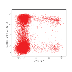
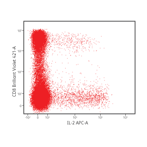
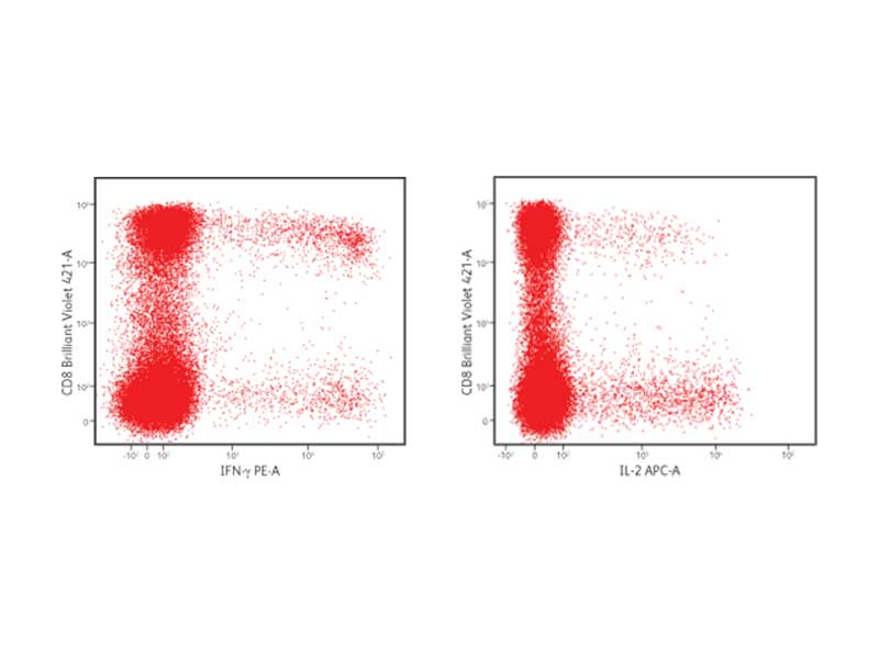
CD8+细胞中的IFN-γ和IL-2分泌。
在存在布雷菲德菌素A的情况下,用葡萄球菌肠毒素B刺激外周血单核细胞6小时。刺激后,使用BD Cytofix/Cytoperm™缓冲系统固定和透化细胞。使用以下荧光抗体染色细胞:CD3 FITC、CD4 PerCP-Cy5.5、CD8 BD Horizon Brilliant Violet™ 421、IFN-γ PE和IL-2 APC。然后使用BD FACSVerse™流式细胞仪对细胞进行清洗和分析。
| Species | Cytokines | Transport Inhibitor |
|---|---|---|
| Human | IL-1α, IL-6, IL-8, TNF-α | Monensin |
| Human | IFN-γ, IL-2, IL-10, IL-12, MCP-1, MCP-3, MIG, MIP-1α, RANTES | Either Monensin or Brefeldin A |
| Mouse | IL-6, IL-12, TNF-α | Brefeldin A |
| Mouse | GM-CSF, IL-3, IL-4, IL-5, IL-10 | Monensin |
| Mouse | IFN-γ, IL-2 | Either Monensin or Brefeldin A |
IFN-γ and IL-2 production in CD8+ cells
PBMCs were stimulated with staphylococcal enterotoxin B for 6 hours in the presence of brefeldin A. After stimulation, cells were fixed and permeabilized using the BD Cytofix/Cytoperm™ Buffer System. Cells were stained with the following fluorescent antibodies: CD3 FITC, CD4 PerCP-Cy5.5, CD8 BD Horizon Brilliant Violet™ 421, IFN-γ PE and IL-2 APC. Cells were then washed and analyzed using the BD FACSVerse™ Flow Cytometer.
BD Biosciences has simplified the detection of cytokines and cell surface markers with kits, buffer systems and rich panels of fluorescent antibodies. Antibodies to surface markers and cytokines conjugated to a variety of fluorochromes are available. This allows for flexibility in staining panel design, supporting high content multicolor flow cytometric analyses to gain the most data from precious cell samples.
BD FastImmune™ Kits contain tested cocktails of fluorescent antibodies, an appropriate protein transport inhibitor, compatible buffers and a detailed protocol for the optimal preparation, staining and detection of cytokine-producing cells from whole blood. Onboard assays on the BD FACSVerse™ Flow Cytometer System further simplify the process.
BD Cytofix/Cytoperm™ Buffer has been cited in thousands of publications for the analysis of cytokine-producing cells by flow cytometry. Researchers select the protein transport inhibitor best suited to their cytokine of interest and use the BD Cytofix/Cytoperm™ Buffer System to fix, permeabilize and stain their cells for flow cytometric analysis.
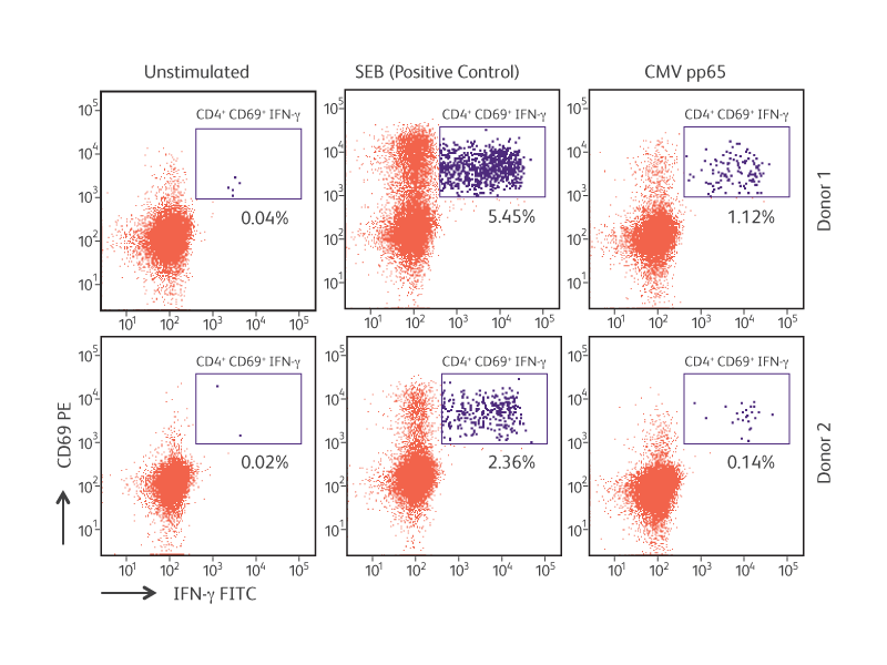
Antigen-specific IFN-γ production by cytomegalovirus (CMV) pp65-stimulated CD4+CD69+ T lymphocytes.
References
- Seder RA, Darrah PA, Roederer M. T-cell quality in memory and protection: implications for vaccine design. Nat Rev Immunol. 2008;8(4):247-258. doi: 10.1038/nri2274
- Schuerwegh AJ, Stevens WJ, Bridts CH, De Clerck LS. Evaluation of monensin and brefeldin A for flow cytometric determination of interleukin-1 beta, interleukin-6, and tumor necrosis factoralpha in monocytes. Cytometry. 2001;46(3):172-176. doi: 10.1002/cyto.1102
BD生物科学公司利用试剂盒、缓冲系统和丰富的荧光抗体流式方案简化了细胞因子和细胞表面标记物的检测程序。可获取结合到各种荧光色素的表面标记物和细胞因子的抗体。这样就保证了染色流式方案设计的灵活性,并支持高含量的多色流式细胞术分析,从而可以从宝贵的细胞样本中获得最多的数据。
BD FastImmune™试剂盒包含经过测试的荧光抗体混合剂、合适的蛋白运输抑制剂、兼容的缓冲液和详细的实验方案,以完成全血细胞因子分泌细胞的最优制备、染色和检测。BD FACSVerse™流式细胞仪系统上的板载分析进一步简化了该过程。
BD Cytofix/Cytoperm™Buffer已被数以千计的出版物引用,用于流式细胞术分析细胞因子分泌细胞。研究人员选择最适合他们所关注细胞因子的蛋白转运抑制剂,并使用BD Cytofix/Cytoperm™缓冲系统来固定、透化和染色他们的细胞,用于流式细胞术分析。
巨细胞病毒(CMV)pp65刺激的CD4+CD69+ T淋巴细胞分泌的抗原特异性IFN-γ
转录因子
细胞内流式细胞术能够检测异质细胞群内的转录因子及其相关蛋白。
同时分析多个标记物可以确定关键的细胞沿着特定的分化途径移动时间点、标记物和频率。
转录因子是与DNA和其他蛋白质结合来调节基因表达的蛋白质。它们在细胞发育和分化中起关键作用。例如,用于分化调节性T细胞的FoxP3以及用于最终内胚层分化的Sox17。
FoxP3被认为是调节性T细胞的主要调控因子。和许多转录因子一样,FoxP3可与数千个基因结合,进而上调或下调调节性T细胞的所需基因表达。3 SATB1(一个基因组组织者)会被FoxP3抑制,阻止T效应细胞因子(包括IL-4和IFN-γ)的诱导4。
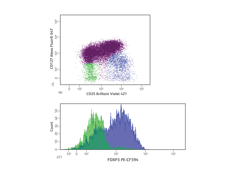
细胞内流式细胞术检测人调节性T细胞。
与细胞内细胞因子染色类似,使用流式细胞术检测转录因子时也需要细胞固定和透化。转录因子通常位于和DNA和其他蛋白质结合的细胞核内。根据靶分子表位的性质,可能需要不同的固定和透化缓冲液。
细胞的固定和透化会影响细胞表面标记物染色,这就使得选择兼容的缓冲液更为关键。为了进一步实现转录因子检测,BD生物科学公司还开发了BD Pharmingen™转录因子缓冲液。该缓冲液能充分透化细胞,暴露核内表位,同时还与许多细胞类型、细胞表面染色和串联染料兼容。
干细胞分化研究便捷试剂盒
当多能干细胞分化为不同的细胞类型时,转录因子和其他蛋白的表达会发生变化。在哺乳动物胚胎发育过程中,定形内胚层会形成肝脏、胰腺和肠道。5,6 在进入最终内胚层的过程中,转录因子Sox17和FoxA2以及细胞表面标记物CD184(CXCR4)的水平会升高,而多能性标记物Nanog和Sox2的水平则下降8
多色流式细胞术是确定表达相关标记物的细胞相对数量的好方法。这对于细胞分化途径的研究,特别是对不同细胞分化方案和分化潜力的优化、量化和比较也是有用的。
为了便于研究干细胞中的转录因子,BD公司开发了几种检测关键干细胞特异性转录因子的试剂盒,包括BD Stemflow™人多能干细胞转录因子分析试剂盒(目录号:560589)、BD Stemflow™小鼠多能干细胞转录因子分析试剂盒(目录号: 560585)、BD Stemflow™人神经细胞谱系分析试剂盒(目录号:561526)以及BD Stemflow™人定形和胰腺内胚层分析试剂盒(目录号:562496)。这些试剂盒包含表征多能干细胞并跟踪多能干细胞分化至各自谱系的抗体和缓冲系统。
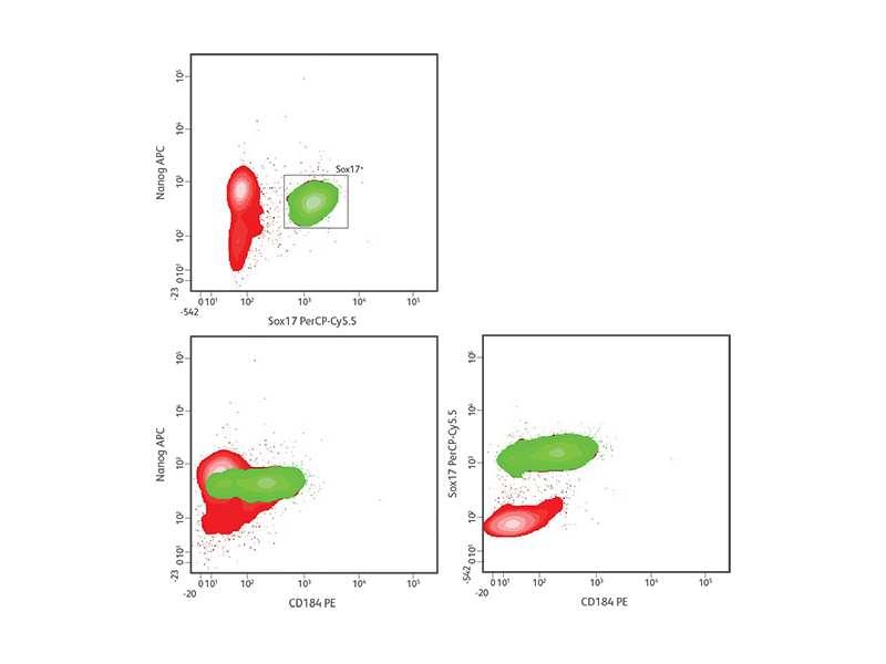
H9造血干细胞的定形内胚层分化。
To facilitate the study of transcription factors in stem cells, BD has developed several kits for the detection of key stem cell–specific transcription factors including the BD Stemflow™ Human Pluripotent Stem Cell Transcription Factor Analysis Kit (Cat. No. 560589), the BD Stemflow™ Mouse Pluripotent Stem Cell Transcription Factor Analysis Kit (Cat. No. 560585), the BD Stemflow™ Human Neural Cell Lineage Analysis Kit (Cat. No. 561526), and the BD Stemflow™ Human Definitive and Pancreatic Endoderm Analysis Kit (Cat. No. 562496). These kits contain antibodies and buffer systems to characterize pluripotent stem cells and track the differentiation of pluripotent stem cells into respective lineages.
References
- Birzele F, Fauti T, Stahl H, et al. Next-generation insights into regulatory T cells: expression profiling and FoxP3 occupancy in Human. Nucleic Acids Res. 2011;39(18):7946-7960. doi: 10.1093/nar/gkr444
- Beyer M, Thabet Y, Müller RU, et al. Repression of the genome organizer SATB1 in regulatory T cells is required for suppressive function and inhibition of effector differentiation. Nat Immunol. 2011;12(9):898-907. doi: 10.1038/ni.2084
- Wang P, McKnight KD, Wong DJ, et al. A molecular signature for purified definitive endoderm guides differentiation and isolation of endoderm from mouse and human embryonic stem cells. Stem Cells Dev. 2012;21(12):2273-2287. doi: 10.1089/scd.2011.0416
- Takayama K, Inamura M, Kawabata K, et al. Efficient and directive generation of two distinct endoderm lineages from human ESCs and iPSCs by differentiation stage-specific SOX17 transduction. PLoS One. 2011;6(7):e21780. doi: 10.1371/journal.pone.0021780
- Murry CE, Keller G. Differentiation of embryonic stem cells to clinically relevant populations: lessons from embryonic development. Cell. 2008;132(4):661-680. doi: 10.1016/j.cell.2008.02.008
- D’Amour KA, Agulnick AD, Eliazer S, Kelly OG, Kroon E, Baetge EE. Efficient differentiation of human embryonic stem cells to definitive endoderm. Nat Biotechnol. 2005;23(12):1534-1541. doi: 10.1038/nbt1163
磷酸化
因为能够在单个细胞的水平上收集数据,流式细胞术在细胞信号研究中有多个优势。
与基于裂解的方法不同,流式细胞术可便于异质信号反应的检测和分析。因此,可以区分一小群细胞内的强大蛋白磷酸化反应与较小但更均匀的反应。通过添加细胞表面标记物的特异性荧光抗体至复杂细胞混合物(如全血)内的细胞亚群,可以检测蛋白磷酸化介导的信号反应。此外,在细胞没有预富集的情况下,也可以发现罕见细胞群。因此,从有限的细胞样本中获得了丰富的数据。
BD Phosflow™产品包括一个优化的缓冲液和荧光单克隆抗体系统,用于流式细胞术检测细胞内信号分子和特定翻译后修饰。为了使用流式细胞术检测特异性磷酸化表位,需固定细胞来维持信号蛋白的磷酸化状态,然后将细胞透化使抗体进入细胞并与靶蛋白特异性结合。然后,对染色细胞的流式细胞术分析可捕获细胞内信号传导和蛋白磷酸化的快照。
蛋白磷酸化本质上是瞬时的,并受到蛋白磷酸酶的严格调控。大多数磷蛋白质检测方法都需要合适的刺激时间点和磷酸酶的迅速失活,包括蛋白质免疫印迹分析。同样,必须快速固定用于流式细胞术分析的细胞样本,以维持磷酸化表位。另一个重要的考虑因素是磷蛋白质的表达水平。可能很难在单细胞水平检测某些蛋白磷酸化活动,特别是当细胞表达低水平的特定信号蛋白或相关位置显示不完全磷酸化时。BD还提供直接与明亮荧光色素结合的高质量抗体,极大地提高了磷蛋白质的细胞内检测。
Multiparameter flow cytometry offers key advantages for the detection and analysis of intracellular signaling within cells and cell subsets in complex mixtures. Since cell signaling studies often examine cellular responses to treatment with stimulatory or inhibitory molecules, unstimulated or untreated cells often provide the best controls for evaluating background staining. Unlike an immunoglobulin isotype control, an unstimulated cell control accounts for the unique background characteristics of each antibody as well as the basal phosphoprotein expression levels within the cells of interest. Cellular permeabilization is required to expose phosphorylated epitopes. Although multiple buffer systems are available, BD Phosflow™ Perm Buffer III is recommended for most applications. The BD Biosciences website contains useful information about the performance of various BD cell surface marker and intracellular antibodies in different buffer conditions and staining protocols.
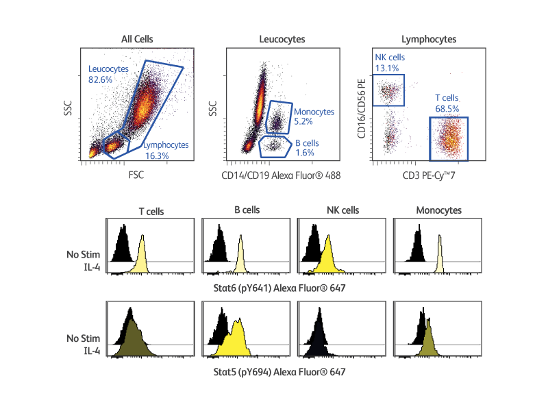
BD Phosflow™ Kits provide useful tools for analyzing the signaling responses elicited by cells of the adaptive and innate immune systems. These kits include fluorescent antibody cocktails specific for cell surface markers to identify specific leucocyte subsets as well as fluorescent antibodies to key phosphoproteins. In addition, the kits provide compatible buffers, positive and negative control cells, and detailed protocols. The BD Phosflow™ Human Monocyte/NK Cell Activation Kit (Cat. No. 562089) allows the simultaneous study of protein phosphorylation in B cells, T cells, monocytes and NK cells from human whole blood, while the BD Phosflow™ Human T-Cell Activation Kit (Cat. No. 560750) is useful for the study of phosphoprotein responses in CD4 versus CD8 T cells.
多参数流式细胞术在检测和分析复杂混合物中的细胞和细胞亚群的细胞内信号传导时具有关键优势。由于细胞信号研究经常检查细胞对刺激性或抑制性分子处理的反应,未刺激或未处理的细胞通常是评价背景染色的最佳对照。与免疫球蛋白同型对照不同,未刺激细胞对照可以解释每种抗体的独特背景特征以及相关细胞内的基础磷蛋白质表达水平。需要通过细胞透化来暴露磷酸化的表位。虽然有多种缓冲系统可以使用,但大多数应用场景都推荐使用BD Phosflow™透化缓冲液III。BD生物科学公司网站包含不同缓冲条件和染色方案下各种BD细胞表面标记物和细胞内抗体的性能方面的有用信息。
Phosflow™试剂盒是分析适应性和先天免疫系统细胞所引发的信号反应的有用工具。这些试剂盒包括识别特定白细胞亚群的细胞表面标记物的特异性荧光抗体混合剂,以及关键磷蛋白质的特异性荧光抗体。此外,该试剂盒还提供兼容的缓冲液、阳性和阴性对照细胞以及详细的实验方案。BD Phosflow™人单核细胞/NK细胞激活试剂盒(目录号:562089)可同时研究人全血中B细胞、T细胞、单核细胞和NK细胞的蛋白磷酸化,而Phosflow™人T细胞激活试剂盒(目录号:560750可用于研究CD4和CD8+T细胞的磷蛋白质反应。
多路复用
虽然其他技术也可以分别测量细胞因子、转录因子或磷酸化蛋白,但细胞内流式细胞术可以在单细胞水平上同时测量多个细胞内标记物。
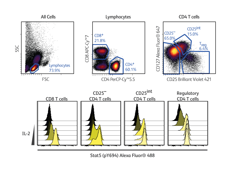
这种方法还能提供信号响应、分化状态和其他细胞活动的数据。结合使用细胞表面和细胞内标记物的特异性荧光抗体可对样品中多种细胞类型的表型和功能差异进行高分辨率比较分析。
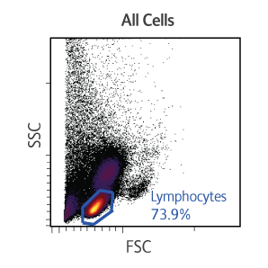
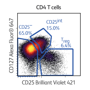
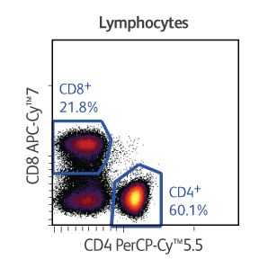
在本例中,在固定、透化并使用表面和细胞内标记物染色之前,先用浓度递增的人重组IL-2刺激低温保存的外周血单核细胞。使用BD Pharmingen™转录因子Phospho Buffer Set可以同时检测对IL-2响应的调节性T细胞中的磷酸化信号转导及转录激活因子5和转录因子FoxP3。
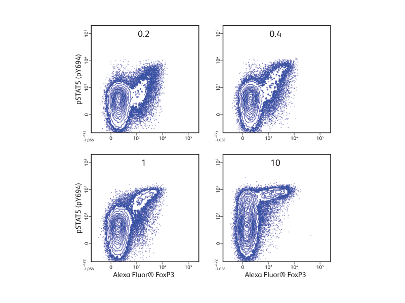
IL-2在T细胞亚群中的剂量反应。
同时测定同一样本内细胞的多种转录因子、细胞因子和表面标记物的表达水平可有效地研究辅助T细胞的分化和功能。RORγt是Th17细胞的标志性转录因子。这对于IL-17的分泌以及CD4+CD8+双阳性胸腺细胞的维持很重要。1,2
在所示实验结果中,细胞分离自BALB/c小鼠胸腺和脾脏。胸腺细胞或脾细胞表面用CD44、CD62L、CD196或适当免疫球蛋白同型对照品的特异性荧光抗体染色。首先用BD Pharmingen™转录因子缓冲液固定和透化细胞,然后用RORγt、Foxp3、IL-17A和IFN-γ特异性荧光抗体进行细胞内染色。
在上图中比较了IL-17A的细胞表达与RORγt、FoxP3(调节性T细胞转录因子)、IFN-γ(Th1细胞因子)的表达。正如预期的那样,胸腺细胞中IL-17A+ RORγt+双阳性细胞较多,而IL-17A与Foxp3或IFN-γ基本没有共表达。脾细胞仅表达了很少的IL-17A。
在下图中,通过进一步分析发现,脾脏中的IL-17A分泌细胞表达表面标记物CD44和CD196(CCR6),但未表达CD62L,这就提供了IL-17A+细胞表型的其他信息。
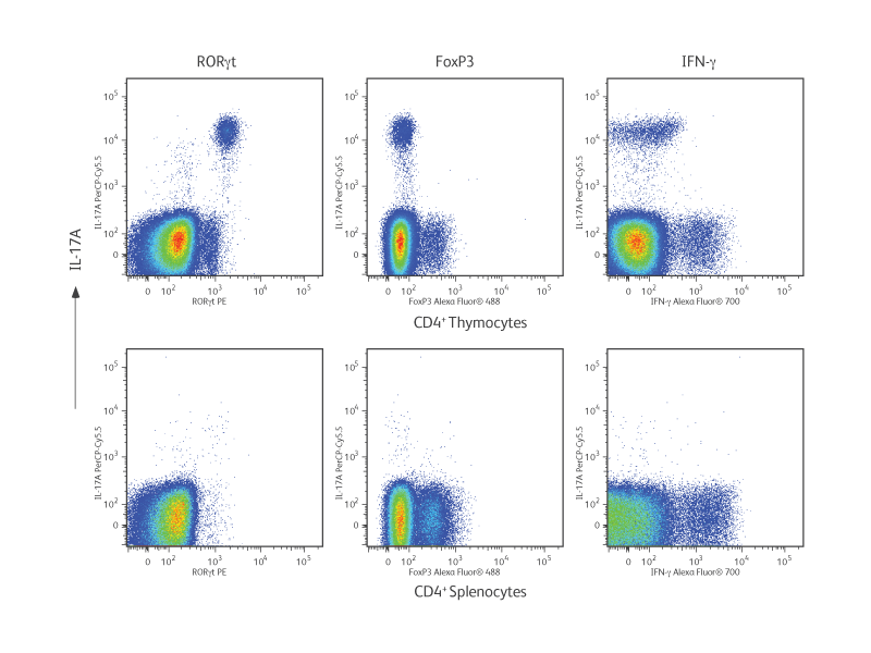
BALB/c胸腺和脾脏Th17细胞的表型分析。
References
- Ghoreschi K, Laurence A, Yang XP, Hirahara K, O’Shea JJ. T helper 17 cell heterogeneity and pathogenicity in autoimmune disease. Trends Immunol. 2011;32(9):395-401. doi: 10.1016/j.it.2011.06.007
- Ivanov II, McKenzie BS, Zhou L, et al. The orphan nuclear receptor RORgammat directs the differentiation program of proinflammatory IL-17+ T helper cells. Cell. 2006;126(6):1121-1133. doi: 10.1016/j.cell.2006.07.035
-
手册

-
常问问题
-
产品信息表
-
方案
-
 96深孔BD Phosflow™细胞因子刺激染色
-
BD Phosflow™96孔深培养板实验方案。
-
Phosflow™替代实验方案1:固定–染色–透化
-
Phosflow™替代实验方案2:染色-固定-透化
-
Phosflow™贴壁细胞实验方案
-
BD Phosflow™人外周血单核细胞实验方案
-
BD Phosflow™人全血样本实验方案
-
BD Phosflow™小鼠脾细胞或胸腺细胞实验方案
-
BD Phosflow™TCR刺激实验方案:人
-
使用BD Horizon™固定活性染料450、BD Cytofix™固定缓冲液和BD Phosflow™透化缓冲液,在解冻和活化的人外周血单核细胞中进行活细胞鉴定并同时进行磷酸化和细胞表面标记物测定。
-
同时测定细胞表面标记物与细胞增殖和蛋白磷酸化。
-
使用BD Phosflow™Lyse/固定缓冲液和BD Phosflow™透化缓冲液III在IL-2刺激的人全血中同时测定T-bet和信号转导及转录激活因子5(pY694)的细胞表面标记物。
-
磷蛋白质检测的建议刺激条件。
-
工具
-
网络研讨会
仅供研究使用,不用于诊疗程序。
Alexa Fluor是Life Technologies Corporation公司的商标。CF是Biotium公司的商标。Cy是全球生命科学解决方案德国股份有限公司的商标,或者是以Cytiva名义开展业务的一家附属公司。Cytobank是美国贝克曼库尔特有限公司的商标。