-
Reagents
- Flow Cytometry Reagents
-
蛋白质印迹试剂
- 免疫分析 试剂
-
Single-Cell Multiomics Reagents
- BD® AbSeq Assay
- BD Rhapsody™ 附件试剂盒
- BD® Single-Cell Multiplexing Kit
- BD Rhapsody™ TCR/BCR Next Multiomic Assays
- BD Rhapsody™ Targeted mRNA Kits
- BD Rhapsody™ Whole Transcriptome Analysis (WTA) Amplification Kit
- BD® OMICS-Guard Sample Preservation Buffer
- BD Rhapsody™ ATAC-Seq Assays
- BD® OMICS-One Protein Panels
-
Functional Assays
-
显微成像试剂
-
Cell Preparation and Separation Reagents
Old Browser
Looks like you're visiting us from {countryName}.
Would you like to stay on the current location or be switched to your location?
BD Pharmingen™ Purified Mouse Anti-Human p53
克隆 PAb 1801 (RUO)


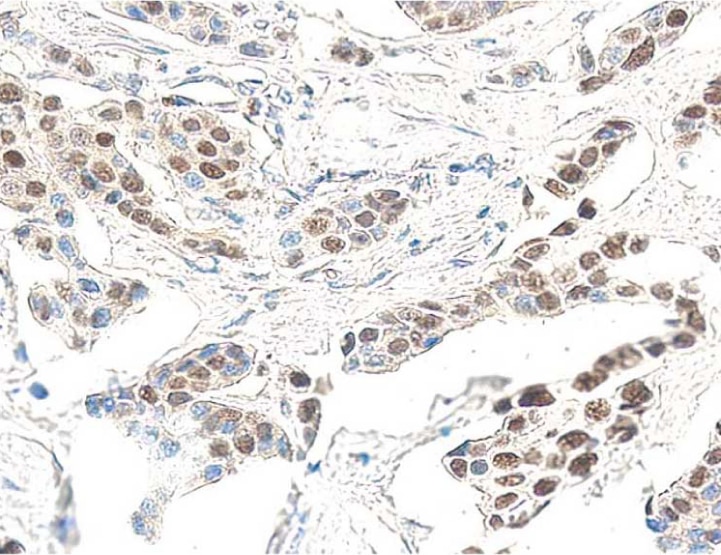
Anti-human p53. Formalin-fixed, paraffin-embedded tissue section of breast carcinoma stained for p53 (clone PAb 1801, Cat. No. 554169) using a DAB chromogen and Hematoxylin counterstain.


监管状态图例
未经BD明确书面授权,严禁使用未经许可的任何商品。
准备和存储
推荐的实验流程
Applications include western blot analysis (1-2 µg/ml), immunoprecipitation (1-2 µg/1 x 10^6 cells), immunofluorescence microscopy of cultured cells, immunohistochemistry of frozen (5-20 µg/ml), and antigen-unmasked paraffin-embedded tissue sections (5-20 µg/ml). Positive control cell lines include SK-BR-3 human breast carcinoma cells (ATCC HTB-30), and A431 human vulval carcinoma cells (ATCC CRL-1555). COS-7 SV40 transformed monkey kidney cells (ATCC CRL-1651) or another SV40-transformed cell line are also useful as positive controls for detecting p53. MCF-7 human breast carcinoma cells (ATCC HTB-22) are suggested as a negative control. Positive immunostaining is seen in a high proportion of breast and colon carcinomas. p53 staining is not typically detected in normal skin, brain, kidney, lung, stomach or breast tissue.
商品通知
- Since applications vary, each investigator should titrate the reagent to obtain optimal results.
- Caution: Sodium azide yields highly toxic hydrazoic acid under acidic conditions. Dilute azide compounds in running water before discarding to avoid accumulation of potentially explosive deposits in plumbing.
- Please refer to www.bdbiosciences.com/us/s/resources for technical protocols.
The gene for the nuclear phosphoprotein p53 is the most commonly mutated gene yet identified in human cancers. Missense mutations occur in tumors of the colon, lung, breast, ovary, bladder and several other organs. The mutant p53 is over-expressed in a variety of transformed cells and it forms specific complexes with several viral oncogenes including SV40 large T, E1B from adenovirus and E6 from human papilloma virus. Recent data suggest that wild type p53 plays a role as a checkpoint protein for DNA damage during the S-phase of the cell cycle. However, it is still unclear whether point mutated forms of p53 are simple null mutants and/or dominant negatively acting proteins. p53 migrates at a reduced molecular weight of 53 kDa. Clone PAb 1801 recognizes an epitope between amino acids 32-79 in the N-terminal domain of human wild type and mutant p53 antibody. It does not cross-react with p53 from other species. A truncated recombinant human p53 fusion protein was used as immunogen.
研发参考 (9)
-
Baker SJ, Markowitz S, Fearon ER, Willson JK, Vogelstein B. Suppression of human colorectal carcinoma cell growth by wild-type p53. Science. 1990; 249(4971):912-915. (Clone-specific: Immunofluorescence). 查看参考
-
Banks L, Matlashewski G, Crawford L. Isolation of human-p53-specific monoclonal antibodies and their use in the studies of human p53 expression. Eur J Biochem. 1986; 159(3):529-534. (Immunogen: Immunoprecipitation, Western blot). 查看参考
-
Jacquemier J, Moles JP, Penault-Llorca F, et al. p53 immunohistochemical analysis in breast cancer with four monoclonal antibodies: comparison of staining and PCR-SSCP results. Br J Cancer. 1994; 69(5):846-852. (Clone-specific: Immunohistochemistry). 查看参考
-
Legros Y, Lacabanne V, d'Agay MF, Larsen CJ, Pla M, Soussi T. Production of human p53 specific monoclonal antibodies and their use in immunohistochemical studies of tumor cells. Bull Cancer. 1993; 80(2):102-110. (Clone-specific: Western blot). 查看参考
-
Porter PL, Gown AM, Kramp SG, Coltrera MD. Widespread p53 overexpression in human malignant tumors. An immunohistochemical study using methacarn-fixed, embedded tissue. Am J Pathol. 1992; 140(1):145-153. (Clone-specific: Immunohistochemistry). 查看参考
-
Said JW, Barrera R, Shintaku IP, Nakamura H, Koeffler HP. Immunohistochemical analysis of p53 expression in malignant lymphomas. Am J Pathol. 1992; 141(6):1343-1348. (Clone-specific: Immunohistochemistry). 查看参考
-
Vogelstein B. Cancer. A deadly inheritance. Nature. 1990; 348(6303):681-682. (Biology). 查看参考
-
Vojtesek B, Bartek J, Midgley CA, Lane DP. An immunochemical analysis of the human nuclear phosphoprotein p53. New monoclonal antibodies and epitope mapping using recombinant p53. J Immunol Methods. 1992; 151(1-2):237-244. (Clone-specific: Immunofluorescence, Immunohistochemistry, Immunoprecipitation, Western blot). 查看参考
-
Walker RA, Dearing SJ, Lane DP, Varley JM. Expression of p53 protein in infiltrating and in-situ breast carcinomas. J Pathol. 1991; 165(3):203-211. (Clone-specific: Immunohistochemistry). 查看参考
Please refer to Support Documents for Quality Certificates
Global - Refer to manufacturer's instructions for use and related User Manuals and Technical data sheets before using this products as described
Comparisons, where applicable, are made against older BD Technology, manual methods or are general performance claims. Comparisons are not made against non-BD technologies, unless otherwise noted.
For Research Use Only. Not for use in diagnostic or therapeutic procedures.