BD Pharmingen™ PE Mouse Anti-Human IgG2
克隆 HP6002 (also known as AB4-DH4) (RUO)

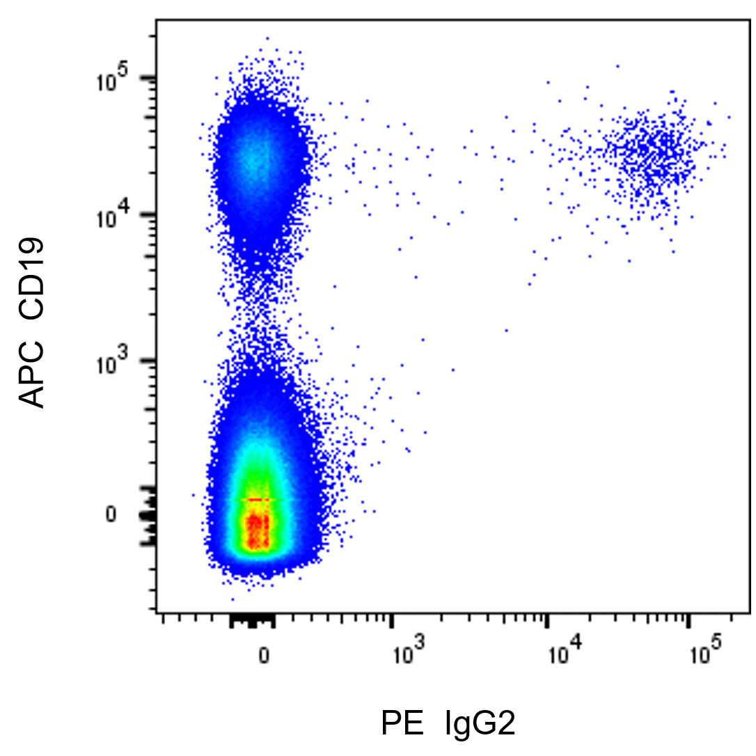
Multicolor flow cytometric analysis of IgG2 expression on Human peripheral blood lymphocytes. Human peripheral blood mononuclear cells (PBMC) were washed and cultured in complete tissue culture medium overnight to minimize subsequent nonspecific immunofluorescent staining. The cells were harvested and stained with APC Mouse Anti-Human CD19 antibody (Cat. No. 555415) and with either PE Mouse IgG1, κ Isotype Control (Cat. No. 349043; Left Plot) or PE Mouse Anti-Human IgG2 antibody (Cat. No. 568959/568960; Right Plot). DAPI (4',6-Diamidino-2-Phenylindole, Dihydrochloride) Solution (Cat. No. 564907) was added to cells right before analysis. The bivariate pseudocolor density plot showing the correlated expression of cell surface IgG2 (or Ig isotype control staining) versus CD19 was derived from gated events with the forward and side light-scatter characteristics of viable (DAPI-negative) lymphocytes. Flow cytometry and data analysis were performed using a BD LSRFortessa™ Cell Analyzer System and FlowJo™ software.
Multicolor flow cytometric analysis of IgG2 expression on Human peripheral blood lymphocytes. Human peripheral blood mononuclear cells (PBMC) were washed and cultured in complete tissue culture medium overnight to minimize subsequent nonspecific immunofluorescent staining. The cells were harvested and stained with APC Mouse Anti-Human CD19 antibody (Cat. No. 555415) and with either PE Mouse IgG1, κ Isotype Control (Cat. No. 349043; Left Plot) or PE Mouse Anti-Human IgG2 antibody (Cat. No. 568959/568960; Right Plot). DAPI (4',6-Diamidino-2-Phenylindole, Dihydrochloride) Solution (Cat. No. 564907) was added to cells right before analysis. The bivariate pseudocolor density plot showing the correlated expression of cell surface IgG2 (or Ig isotype control staining) versus CD19 was derived from gated events with the forward and side light-scatter characteristics of viable (DAPI-negative) lymphocytes. Flow cytometry and data analysis were performed using a BD LSRFortessa™ Cell Analyzer System and FlowJo™ software.
Multicolor flow cytometric analysis of IgG2 expression on Human peripheral blood lymphocytes. Human peripheral blood mononuclear cells (PBMC) were washed and cultured in complete tissue culture medium overnight to minimize subsequent nonspecific immunofluorescent staining. The cells were harvested and stained with APC Mouse Anti-Human CD19 antibody (Cat. No. 555415) and with either PE Mouse IgG1, κ Isotype Control (Cat. No. 349043; Left Plot) or PE Mouse Anti-Human IgG2 antibody (Cat. No. 568959/568960; Right Plot). DAPI (4',6-Diamidino-2-Phenylindole, Dihydrochloride) Solution (Cat. No. 564907) was added to cells right before analysis. The bivariate pseudocolor density plot showing the correlated expression of cell surface IgG2 (or Ig isotype control staining) versus CD19 was derived from gated events with the forward and side light-scatter characteristics of viable (DAPI-negative) lymphocytes. Flow cytometry and data analysis were performed using a BD LSRFortessa™ Cell Analyzer System and FlowJo™ software.
Multicolor flow cytometric analysis of IgG2 expression on Human peripheral blood lymphocytes. Human peripheral blood mononuclear cells (PBMC) were washed and cultured in complete tissue culture medium overnight to minimize subsequent nonspecific immunofluorescent staining. The cells were harvested and stained with APC Mouse Anti-Human CD19 antibody (Cat. No. 555415) and with either PE Mouse IgG1, κ Isotype Control (Cat. No. 349043; Left Plot) or PE Mouse Anti-Human IgG2 antibody (Cat. No. 568959/568960; Right Plot). DAPI (4',6-Diamidino-2-Phenylindole, Dihydrochloride) Solution (Cat. No. 564907) was added to cells right before analysis. The bivariate pseudocolor density plot showing the correlated expression of cell surface IgG2 (or Ig isotype control staining) versus CD19 was derived from gated events with the forward and side light-scatter characteristics of viable (DAPI-negative) lymphocytes. Flow cytometry and data analysis were performed using a BD LSRFortessa™ Cell Analyzer System and FlowJo™ software.
准备和存储
推荐的实验流程
BD® CompBeads can be used as surrogates to assess fluorescence spillover (compensation). When fluorochrome conjugated antibodies are bound to BD® CompBeads, they have spectral properties very similar to cells. However, for some fluorochromes there can be small differences in spectral emissions compared to cells, resulting in spillover values that differ when compared to biological controls. It is strongly recommended that when using a reagent for the first time, users compare the spillover on cell and BD® CompBeads to ensure that BD® CompBeads are appropriate for your specific cellular application.
商品通知
- Please refer to www.bdbiosciences.com/us/s/resources for technical protocols.
- This reagent has been pre-diluted for use at the recommended Volume per Test. We typically use 1 × 10^6 cells in a 100-µl experimental sample (a test).
- An isotype control should be used at the same concentration as the antibody of interest.
- Caution: Sodium azide yields highly toxic hydrazoic acid under acidic conditions. Dilute azide compounds in running water before discarding to avoid accumulation of potentially explosive deposits in plumbing.
- For fluorochrome spectra and suitable instrument settings, please refer to our Multicolor Flow Cytometry web page at www.bdbiosciences.com/colors.
- Please refer to http://regdocs.bd.com to access safety data sheets (SDS).
- Source of all serum proteins is from USDA inspected abattoirs located in the United States.
配套商品


The HP6002 monoclonal antibody specifically recognizes the subclass of human Immunoglobulin G (IgG) known as IgG2. After human IgG1, human IgG2 is the second most abundant of the four subclasses of IgG found in human serum followed by IgG3 and IgG4. IgG2 is comprised of two identical heavy chains encoded by IGHG2, and two light chains, either Igκ or Igλ, linked by disulfide bonds. The HP6002 antibody binds to the CH2 domain of the IgG2 heavy chain and does not crossreact with other immunoglobulin heavy chain (IgH) subclasses. IgG2 is normally expressed by plasmablasts, plasma cells, and memory B cells as well as by some myeloma or plasmacytoma cells. HP6002 binds to soluble human IgG2, cytophilic IgG2 attached to cells through its Fc region, or human IgG2 antibodies specifically bound to antigens. IgG2 can cross the placenta and disseminate throughout extravascular fluids throughout the body. Human IgG2 serves multiple functions with the transmembrane form serving as an antigen receptor for B lymphocytes and secreted soluble forms participating in various effector functions including antibody-dependent neutralization of toxins or infection by microbes and complement fixation.

研发参考 (5)
-
Blanco E, Perez-Andres M, Sanoja-Flores L, et al. Selection and validation of antibody clones against IgG and IgA subclasses in switched memory B-cells and plasma cells.. J Immunol Methods. 2019; 475:112372. (Clone-specific: Flow cytometry). 查看参考
-
Gao B, Moore C, Porcheray F, et al. Pretransplant IgG reactivity to apoptotic cells correlates with late kidney allograft loss.. Am J Transplant. 2014; 14(7):1581-91. (Clone-specific: Flow cytometry). 查看参考
-
Jefferis R, Reimer CB, Skvaril F, et al. Evaluation of monoclonal antibodies having specificity for human IgG sub-classes: results of an IUIS/WHO collaborative study.. Immunol Lett. 1985; 10(3-4):223-52. (Clone-specific: ELISA, Hemagglutination assay, Immunocytochemistry, Immunofluorescence, Radioimmunoassay). 查看参考
-
Reimer CB, Phillips DJ, Aloisio CH, et al. Evaluation of thirty-one mouse monoclonal antibodies to human IgG epitopes.. Hybridoma. 1984; 3(3):263-75. (Immunogen: Immunofluorescence, Immunoprecipitation). 查看参考
-
Tangye SG, Ferguson A, Avery DT, Ma CS, Hodgkin PD. Isotype switching by human B cells is division-associated and regulated by cytokines.. J Immunol. 2002; 169(8):4298-306. (Clone-specific: Flow cytometry). 查看参考
Please refer to Support Documents for Quality Certificates
Global - Refer to manufacturer's instructions for use and related User Manuals and Technical data sheets before using this products as described
Comparisons, where applicable, are made against older BD Technology, manual methods or are general performance claims. Comparisons are not made against non-BD technologies, unless otherwise noted.
For Research Use Only. Not for use in diagnostic or therapeutic procedures.