BD Pharmingen™ PE Mouse Anti-Flavivirus Envelope Protein
克隆 4G2.rMAb (also known as 4G2; D1-4G2-4-15) (RUO)
准备和存储
推荐的实验流程
Bead-based compensation or unmixing controls, such as BD® CompBeads or BD™ SpectraComp™, can be used as surrogates to assess fluorescence spillover when bound to fluorochrome-conjugated antibodies. Although these beads have spectral properties similar to cells, variations in spectral emission may occur, resulting in differing spillover values compared to biological controls. Therefore, it is considered best practice to compare the spillover obtained from cells and bead-based controls when using BD® CompBeads or BD™ SpectraComp™ for the first time, to ensure they are appropriate for the intended application.
商品通知
- Please refer to www.bdbiosciences.com/us/s/resources for technical protocols.
- Caution: Sodium azide yields highly toxic hydrazoic acid under acidic conditions. Dilute azide compounds in running water before discarding to avoid accumulation of potentially explosive deposits in plumbing.
- Since applications vary, each investigator should titrate the reagent to obtain optimal results.
- For fluorochrome spectra and suitable instrument settings, please refer to our Multicolor Flow Cytometry web page at www.bdbiosciences.com/colors.
- An isotype control should be used at the same concentration as the antibody of interest.
- Please refer to http://regdocs.bd.com to access safety data sheets (SDS).
- For U.S. patents that may apply, see bd.com/patents.
配套商品


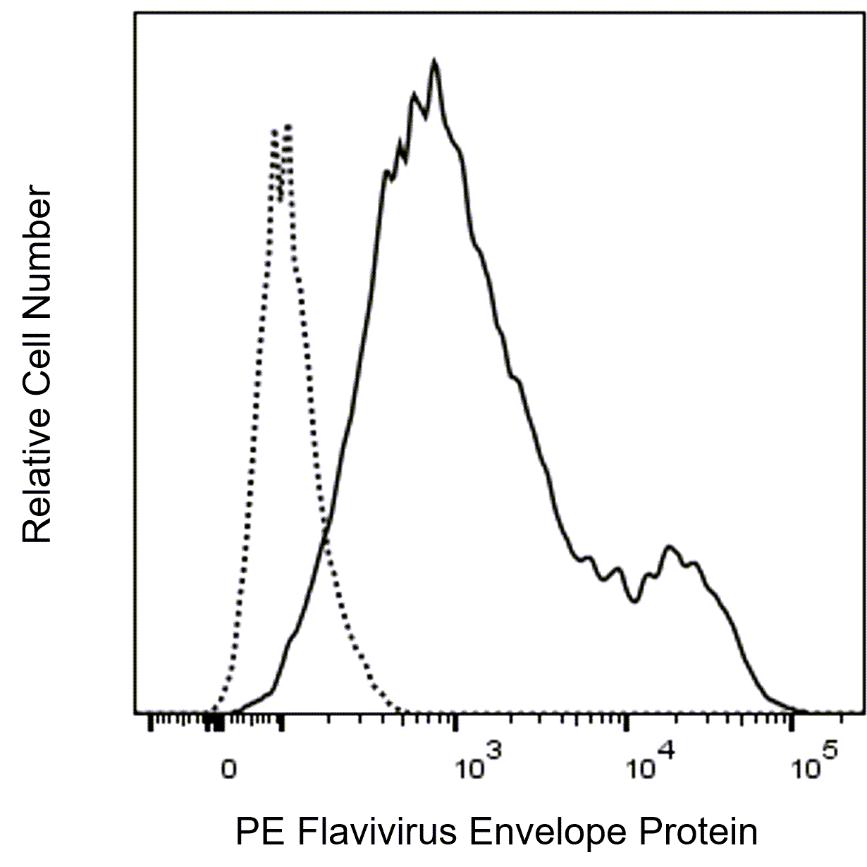
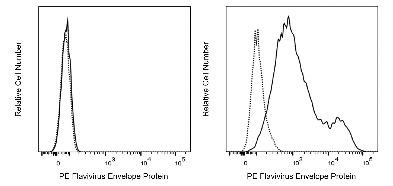
The 4G2 recombinant monoclonal antibody specifically recognizes the Flavivirus Envelope Protein, which is also known as Flavivirus E Protein or E Glycoprotein. This multifunctional class II fusion protein, structurally located on the outermost side of the virion, is divided into three structural envelope domains (DI, DII and DIII), a helical stem, and a transmembrane domain. Its intricate domains contribute to viral entry, fusion, and immune recognition. Cell entry is facilitated by a conserved peptide of 16 amino acids located in the DII region of the Flavivirus Envelope Protein, which includes the immunodominant fusion loop epitope, making it a critical target for antiviral research. The 4G2 antibody reportedly binds to the E protein of flavivirus group-specific antigens, such as Dengue Virus (DENV), Zika Virus (ZIKV), West Nile Virus (WNV), Japanese Encephalitis Virus (JEV) and Yellow Fever Virus (YFV), among others.

研发参考 (9)
-
Gentry MK, Henchal EA, McCown JM, Brandt WE, Dalrymple JM. Identification of distinct antigenic determinants on dengue-2 virus using monoclonal antibodies.. Am J Trop Med Hyg. 1982; 31(3 Pt 1):548-55. (Immunogen: Intracellular Staining/Flow Cytometry). 查看参考
-
Hu T, Wu Z, Wu S, Chen S, Cheng A. The key amino acids of E protein involved in early flavivirus infection: viral entry.. Virol J. 2021; 18(1):136. (Biology). 查看参考
-
Kaufusi PH, Tseng AC, Kelley JF, Nerurkar VR. Selective Reactivity of Anti-Japanese Encephalitis Virus NS4B Antibody Towards Different Flaviviruses.. Viruses. 2020; 12(2):212. (Clone-specific: Immunofluorescence, Western blot). 查看参考
-
Lannes N, Garcia-Nicolàs O, Démoulins T, Summerfield A, Filgueira L. CX3CR1-CX3CL1-dependent cell-to-cell Japanese encephalitis virus transmission by human microglial cells.. Sci Rep. 2019; 9(1):4833. (Clone-specific). 查看参考
-
Martins ST, Kuczera D, Lötvall J, Bordignon J, Alves LR. Characterization of Dendritic Cell-Derived Extracellular Vesicles During Dengue Virus Infection.. Front Microbiol. 2018; 9:1792. (Clone-specific: Intracellular Staining/Flow Cytometry). 查看参考
-
Ricciardi-Jorge T, Bordignon J, Koishi A, Zanluca C, Mosimann AL, Duarte Dos Santos CN. Development of a quantitative NS1-capture enzyme-linked immunosorbent assay for early detection of yellow fever virus infection.. Sci Rep. 2017; 7(1):16229. (Clone-specific: Intracellular Staining/Flow Cytometry). 查看参考
-
Riedl W, Acharya D, Lee JH, et al. Zika Virus NS3 Mimics a Cellular 14-3-3-Binding Motif to Antagonize RIG-I- and MDA5-Mediated Innate Immunity.. Cell Host Microbe. 2019; 26(4):493-503.e6. (Clone-specific: Intracellular Staining/Flow Cytometry). 查看参考
-
Shigeta K, Hayashida T, Hoshino Y, et al. Expression of Epidermal Growth Factor Receptor Detected by Cetuximab Indicates Its Efficacy to Inhibit In Vitro and In Vivo Proliferation of Colorectal Cancer Cells.. PLoS One. 2013; 8(6):e66302. (Clone-specific: Immunofluorescence, Intracellular Staining/Flow Cytometry). 查看参考
-
de Freitas CS, Higa LM, Sacramento CQ, et al. Yellow fever virus is susceptible to sofosbuvir both in vitro and in vivo.. PLoS Negl Trop Dis. 2019; 13(1):e0007072. (Clone-specific: Intracellular Staining/Flow Cytometry). 查看参考
Please refer to Support Documents for Quality Certificates
Global - Refer to manufacturer's instructions for use and related User Manuals and Technical data sheets before using this products as described
Comparisons, where applicable, are made against older BD Technology, manual methods or are general performance claims. Comparisons are not made against non-BD technologies, unless otherwise noted.
For Research Use Only. Not for use in diagnostic or therapeutic procedures.