-
Reagents
- Flow Cytometry Reagents
-
蛋白质印迹试剂
- 免疫分析 试剂
-
Single-Cell Multiomics Reagents
- BD® AbSeq Assay
- BD Rhapsody™ 附件试剂盒
- BD® Single-Cell Multiplexing Kit
- BD Rhapsody™ Targeted mRNA Kits
- BD Rhapsody™ Whole Transcriptome Analysis (WTA) Amplification Kit
- BD Rhapsody™ TCR/BCR Profiling Assays
- BD® OMICS-Guard Sample Preservation Buffer
- BD Rhapsody™ ATAC-Seq Assays
- BD Rhapsody™ TCR/BCR Next Multiomic Assays
-
Functional Assays
-
显微成像试剂
-
Cell Preparation and Separation Reagents
-
- BD® AbSeq Assay
- BD Rhapsody™ 附件试剂盒
- BD® Single-Cell Multiplexing Kit
- BD Rhapsody™ Targeted mRNA Kits
- BD Rhapsody™ Whole Transcriptome Analysis (WTA) Amplification Kit
- BD Rhapsody™ TCR/BCR Profiling Assays
- BD® OMICS-Guard Sample Preservation Buffer
- BD Rhapsody™ ATAC-Seq Assays
- BD Rhapsody™ TCR/BCR Next Multiomic Assays
- China (Chinese)
-
更改国家/语言
Old Browser
Looks like you're visiting us from {countryName}.
Would you like to stay on the current country site or be switched to your country?


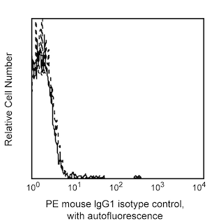
Flow cytometric analysis of CD151 expression on human peripheral blood platelets. Platelets were stained with either PE Mouse Anti-Human CD151 (Cat. No. 556057; solid line histogram) or PE Mouse IgG1, κ Isotype Control (Cat. No. 555749; dashed line histogram). Fluorescent histograms were derived from gated events with the side and forward light-scattering characteristics of viable platelets.


BD Pharmingen™ PE Mouse Anti-Human CD151

监管状态图例
未经BD明确书面授权,严禁使用未经许可的任何商品。
准备和存储
商品通知
- This reagent has been pre-diluted for use at the recommended Volume per Test. We typically use 1 × 10^6 cells in a 100-µl experimental sample (a test).
- An isotype control should be used at the same concentration as the antibody of interest.
- Caution: Sodium azide yields highly toxic hydrazoic acid under acidic conditions. Dilute azide compounds in running water before discarding to avoid accumulation of potentially explosive deposits in plumbing.
- Source of all serum proteins is from USDA inspected abattoirs located in the United States.
- For fluorochrome spectra and suitable instrument settings, please refer to our Multicolor Flow Cytometry web page at www.bdbiosciences.com/colors.
- Please refer to www.bdbiosciences.com/us/s/resources for technical protocols.
Reacts with platelet-endothelial cell tetraspan antigen-3 (PETA-3), a 27 kD membrane glycoprotein, expressed on platelets, megakaryocytes, lymphocytes (weak), monocytes, endothelial cells and epithelial cells. PETA-3 (CD151) associates with β1 integrin in certain tissues. This has also been shown with other tetraspan superfamily members, like CD9, CD63 and α5β1. Reports indicate that this association or colocalization of CD151 with β1 integrin in tissues suggests a functional role of this molecule, however, this role has not been elucidated yet. It has also been reported that antibody 14A2.H1 is capable of platelet activation in vitro. Studies showed that different clones of CD151 monoclonal antibodies display strikingly different patterns of binding to human haemopoietic cells and tissue sections, and that this is due at least in part to the presence of the protein in complexes with different integrins.

研发参考 (5)
-
Fitter S, Tetaz TJ, Berndt MC, Ashman LK. Molecular cloning of cDNA encoding a novel platelet-endothelial cell tetra-span antigen, PETA-3. Blood. 1995; 86(4):1348-1355. (Biology). 查看参考
-
Geary SM, Cambareri AC, Sincock PM, Fitter S, Ashman LK. Differential tissue expression of epitopes of the tetraspanin CD151 recognised by monoclonal antibodies.. Tissue Antigens. 2001; 58(3):141-53. (Clone-specific: Flow cytometry, Immunohistochemistry, Immunoprecipitation). 查看参考
-
Kishimoto T. Tadamitsu Kishimoto .. et al., ed. Leucocyte typing VI : white cell differentiation antigens : proceedings of the sixth international workshop and conference held in Kobe, Japan, 10-14 November 1996. New York: Garland Pub.; 1997.
-
Roberts JJ, Rodgers SE, Drury J, Ashman LK, Lloyd JV. Platelet activation induced by a murine monoclonal antibody directed against a novel tetra-span antigen. Br J Haematol. 1995; 89(4):853-860. (Biology). 查看参考
-
Sincock PM, Mayrhofer G, Ashman LK. Localization of the transmembrane 4 superfamily (TM4SF) member PETA-3 (CD151) in normal human tissues: comparison with CD9, CD63, and alpha5beta1 integrin. J Histochem Cytochem. 1997; 45(4):515-525. (Biology). 查看参考
Please refer to Support Documents for Quality Certificates
Global - Refer to manufacturer's instructions for use and related User Manuals and Technical data sheets before using this products as described
Comparisons, where applicable, are made against older BD Technology, manual methods or are general performance claims. Comparisons are not made against non-BD technologies, unless otherwise noted.
For Research Use Only. Not for use in diagnostic or therapeutic procedures.
Report a Site Issue
This form is intended to help us improve our website experience. For other support, please visit our Contact Us page.
