-
Reagents
- Flow Cytometry Reagents
-
蛋白质印迹试剂
- 免疫分析 试剂
-
Single-Cell Multiomics Reagents
- BD® AbSeq Assay
- BD Rhapsody™ 附件试剂盒
- BD® Single-Cell Multiplexing Kit
- BD Rhapsody™ TCR/BCR Next Multiomic Assays
- BD Rhapsody™ Targeted mRNA Kits
- BD Rhapsody™ Whole Transcriptome Analysis (WTA) Amplification Kit
- BD OMICS-Guard™ Sample Preservation Buffer
- BD Rhapsody™ ATAC-Seq Assays
- BD® OMICS-One Protein Panels
- BD OMICS-One™ WTA Next Assay
-
Functional Assays
-
显微成像试剂
-
Cell Preparation and Separation Reagents
Old Browser
Looks like you're visiting us from {countryName}.
Would you like to stay on the current location or be switched to your location?
BD Horizon™ BUV395 Rat Anti-Mouse F4/80
克隆 T45-2342 (RUO)
Two-color flow cytometric analysis of F4/80 expression on mouse splenocytes. C57BL/6 mouse splenic leucocytes were preincubated with Purified Rat Anti-Mouse CD16/CD32 antibody (Mouse BD Fc Block™) (Cat. No. 553141/553142) and stained with APC Rat Anti-Mouse CD11b antibody (Cat. No. 553312/561690) and either BD Horizon™ BUV395 Rat IgG2a Isotype Control (Cat. No. 563556; Left Plot) or BD Horizon BUV395 Rat Anti-Mouse F4/80 antibody (Cat. No. 565614; Right Plot). The two-color flow cytometric dot plot showing the correlated expression of F4/80 (or Ig Isotype control staining) versus CD11b was derived from gated events with the forward and side light-scatter characteristics of viable monocytes. Flow cytometric analyses were performed using a BD LSRFortessa™ Cell Analyzer System.
Two-color flow cytometric analysis of F4/80 expression on mouse peritoneal cells. C57BL/6 mouse peritoneal cells were preincubated with Mouse BD Fc Block™ and stained with APC Rat Anti-Mouse CD117 (Cat. No. 553356/561074) and BD Horizon BUV395 Rat Anti-Mouse F4/80 antibodies. The two-color dot plot showing the correlated expression of F4/80 versus CD117 was derived from gated events with the light-scatter characteristics of viable peritoneal cells. Flow cytometric analyses were performed using a BD LSRFortessa™ Cell Analyzer System.
监管状态图例
未经BD明确书面授权,严禁使用未经许可的任何商品。
准备和存储
推荐的实验流程
For optimal and reproducible results, BD Horizon Brilliant™ Stain Buffer should be used anytime BD Horizon Brilliant™ dyes are used in a multicolor flow cytometry panel. Fluorescent dye interactions may cause staining artifacts which may affect data interpretation. The BD Horizon Brilliant Stain Buffer was designed to minimize these interactions. When BD Horizon Brilliant Stain Buffer is used in in the multicolor panel, it should also be used in the corresponding compensation controls for all dyes to achieve the most accurate compensation. For the most accurate compensation, compensation controls created with either cells or beads should be exposed to BD Horizon Brilliant Stain Buffer for the same length of time as the corresponding multicolor panel. More information can be found in the Technical Data Sheet of the BD Horizon Brilliant Stain Buffer (Cat. No. 563794/566349) or the BD Horizon Brilliant Stain Buffer Plus (Cat. No. 566385).
商品通知
- Since applications vary, each investigator should titrate the reagent to obtain optimal results.
- Please refer to www.bdbiosciences.com/us/s/resources for technical protocols.
- Caution: Sodium azide yields highly toxic hydrazoic acid under acidic conditions. Dilute azide compounds in running water before discarding to avoid accumulation of potentially explosive deposits in plumbing.
- For fluorochrome spectra and suitable instrument settings, please refer to our Multicolor Flow Cytometry web page at www.bdbiosciences.com/colors.
- Source of all serum proteins is from USDA inspected abattoirs located in the United States.
- An isotype control should be used at the same concentration as the antibody of interest.
配套商品
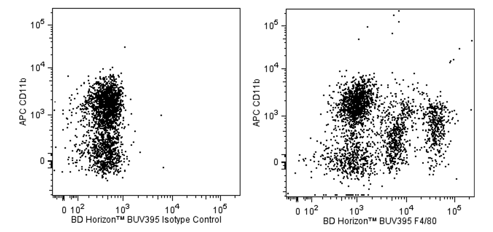
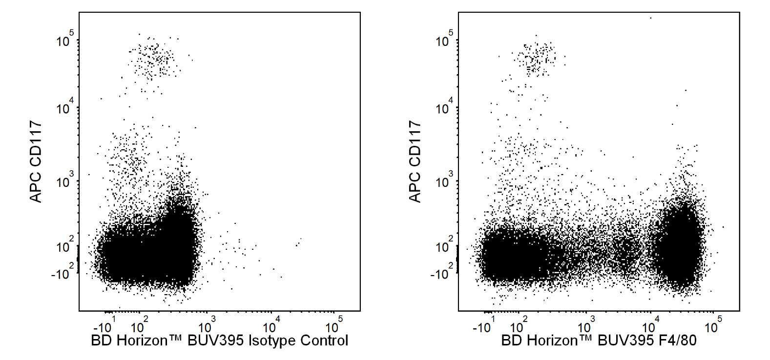
The T45-2342 monoclonal antibody recognizes the mouse F4/80 antigen which is also known as EGF-like module-containing mucin-like hormone receptor-like 1 (EMR1). F4/80 is a 160 kDa glycoprotein that belongs to the EGF-TM7 family of seven-transmembrane spanning cell surface molecules. It is expressed on the surface of granulocytes and a wide range of mature tissue macrophages including, Kupffer cells, splenic red pulp macrophages, microglia, gut lamina propria macrophages, and Langerhans cells. F4/80 expression has also been reported on subpopulations of dendritic cells. F4/80 expression is heterogeneous and may be increased during inflammatory responses as observed in various mouse models of colitis, diabetes and brain injury.
研发参考 (6)
-
Austyn JM., and Gordon S. F4/80, a monoclonal antibody directed specifically against the mouse macrophage. Eur J Immunol. 1981; 10:805-815. (Biology). 查看参考
-
Bodhankar S, Lapato A, Chen Y, Vandenbark AA, Saugstad JA, Offner H. Role for microglia in sex differences after ischemic stroke: importance of M2.. Metab Brain Dis. 2015. (Clone-specific: Flow cytometry). 查看参考
-
Gordon S, Hamann J, Lin HH, Stacey M. F4/80 and the related adhesion-GPCRs. Eur J Immunol. 2011; 41(9):2472-2476. (Biology). 查看参考
-
Krüger T, Benke D, Eitner F, et al. Identification and functional characterization of dendritic cells in the healthy murine kidney and in experimental glomerulonephritis. J Am Soc Nephrol. 2004; 15(3):613-621. (Biology). 查看参考
-
Leenen PJ, Radosević K, Voerman JS, et al. Heterogeneity of mouse spleen dendritic cells: in vivo phagocytic activity, expression of macrophage markers, and subpopulation turnover.. J Immunol. 1998; 160(5):2166-73. (Biology). 查看参考
-
McKnight AJ, Macfarlane AJ, Dri P, Turley L, Willis AC, Gordon S. Molecular cloning of F4/80, a murine macrophage-restricted cell surface glycoprotein with Homology to the G-protein-linked transmembrane & hormone receptor family. J Biol Chem. 1996; 271:486. (Biology). 查看参考
Please refer to Support Documents for Quality Certificates
Global - Refer to manufacturer's instructions for use and related User Manuals and Technical data sheets before using this products as described
Comparisons, where applicable, are made against older BD Technology, manual methods or are general performance claims. Comparisons are not made against non-BD technologies, unless otherwise noted.
For Research Use Only. Not for use in diagnostic or therapeutic procedures.