Old Browser
Looks like you're visiting us from {countryName}.
Would you like to stay on the current country site or be switched to your country?


Immunoprecipitiation/western blot analysis of Cdk4. HeLa or 293 cell lysates were immunoprecipitated with an antibody to Cdk4 (clone DCS-156). Proteins were separated by SDS/PAGE and then western blotted with clone DCS-156. The bands above and below the ~32 kD Cdk4 bands, represent the heavy and light chains of IgG used for immunoprecipitation.

Western blot titration of anti-Cdk4 antibody. HeLa or 293 cell lysates were probed with 6 ug/ml (lanes 1 and 4), 2.0 µg/ml (lanes 2 and 4) or 0.5 µg/ml (lanes 3 and 5) of antibody (clone DCS-156). The antibody identifies Cdk4 at ~32 kD.


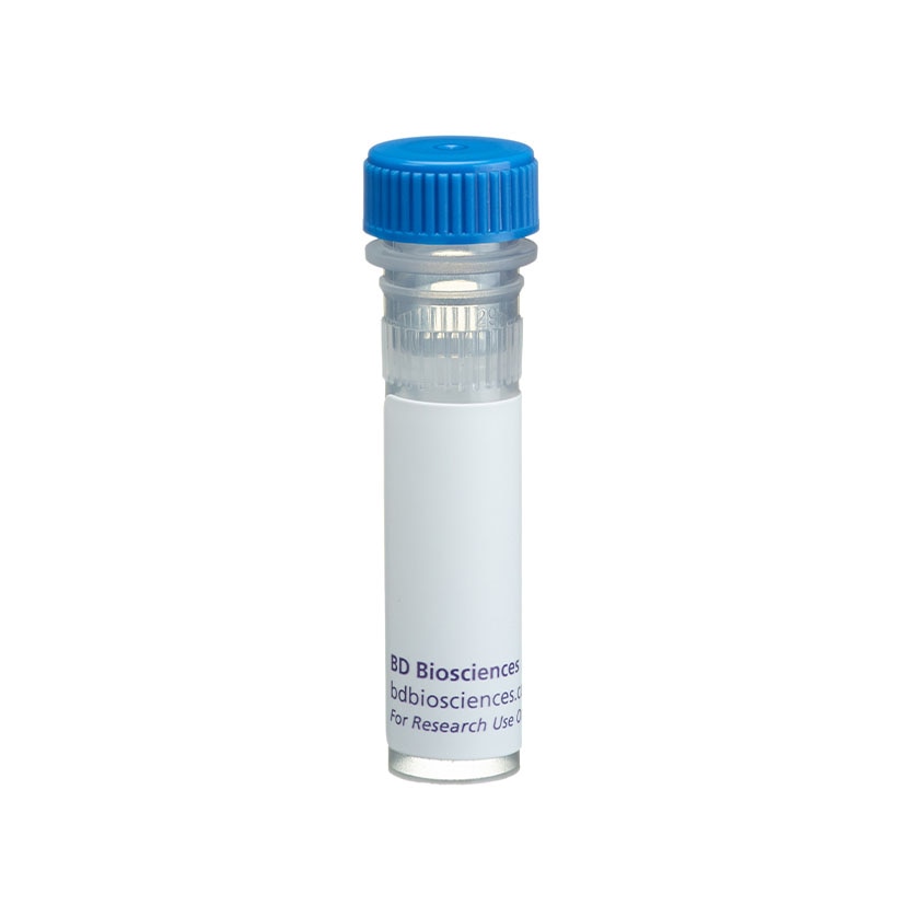
BD Pharmingen™ Purified Mouse Anti-Cdk4

BD Pharmingen™ Purified Mouse Anti-Cdk4

监管状态图例
未经BD明确书面授权,严禁使用未经许可的任何商品。
准备和存储
推荐的实验流程
Applications include western blot analysis (0.5-2.0 µg/ml) and immunoprecipitation (1.0 µg/one million cells). The antibody has also been used for immunohistochemistry of formalin-fixed, paraffin-embedded tissue sections, but this application is not routinely tested at BD Biosciences Pharmingen. HeLa (ATCC CCL 2), 293 (ATCC CRL 1573), or NIH/3T3 (ATCC CRL 1658) cells are suggested as positive controls.
商品通知
- Since applications vary, each investigator should titrate the reagent to obtain optimal results.
- Please refer to www.bdbiosciences.com/us/s/resources for technical protocols.
- Caution: Sodium azide yields highly toxic hydrazoic acid under acidic conditions. Dilute azide compounds in running water before discarding to avoid accumulation of potentially explosive deposits in plumbing.
Cyclins, cyclin-dependent kinases (Cdks), and cyclin-dependent kinase inhibitors (CdkIs) are essential for cell-cycle control in eukarytotes. Cyclins, regulatory subunits, bind to cyclin-dependent kinases (Cdks), catalytic subunits, to form active cyclin-Cdk complexes. Cdk subunits by themselves are inactive and binding to a cyclin is required for their activity. Cyclins A, B1, D and E undergo periodic synthesis and degradation, thereby providing a mechanism to regulate Cdk activity throughout the cell cycle. Additionally, Cdk activity is further regulated by activating and inhibitory phosphorylations, and small proteins (p15, p16, p18, p19, p21 and p27), called inhibitors of Cdk activity, that bind to cyclins, Cdks, or cyclin- Cdk complexes. Cdk4 was originally called PSK-J3, and following demonstration of its association with D-type cyclins, became known as Cdk4. D-type cyclins also associate with Cdks 2 and 5, although Cdk4 appears to be the most abundant partner. The D-type cyclins (D1, D2, and D3) are expressed in response to growth factors or mitogens, and rapidly degrade when mitogens are withdrawn. D cyclins appear to promote G0 to G1 transitions and the rate of G1 progression. For example, cyclin D-Cdk4 and cyclin D-Cdk6 complexes phosphorylate the retinoblastoma protein (Rb) which removes the G1 phase block caused by underphosphorylated Rb. Cdk4 has a molecular weight of ~32 kD. Clone DCS-156 recognizes human and mouse Cdk4. A recombinant protein fragment from the C-terminal end of human Cdk4 was used as immunogen.
研发参考 (2)
-
Johnson DG, Walker CL. Cyclins and cell cycle checkpoints. Annu Rev Pharmacol Toxicol. 1999; 39:295-312. (Biology). 查看参考
-
Matsushime H, Ewen ME, Strom DK, et al. Identification and properties of an atypical catalytic subunit (p34PSK-J3/cdk4) for mammalian D type G1 cyclins. Cell. 1992; 71(2):323-334. (Biology). 查看参考
Please refer to Support Documents for Quality Certificates
Global - Refer to manufacturer's instructions for use and related User Manuals and Technical data sheets before using this products as described
Comparisons, where applicable, are made against older BD Technology, manual methods or are general performance claims. Comparisons are not made against non-BD technologies, unless otherwise noted.
For Research Use Only. Not for use in diagnostic or therapeutic procedures.
Report a Site Issue
This form is intended to help us improve our website experience. For other support, please visit our Contact Us page.

