-
Reagents
- Flow Cytometry Reagents
-
蛋白质印迹试剂
- 免疫分析 试剂
-
Single-Cell Multiomics Reagents
- BD® AbSeq Assay
- BD Rhapsody™ 附件试剂盒
- BD® Single-Cell Multiplexing Kit
- BD Rhapsody™ Targeted mRNA Kits
- BD Rhapsody™ Whole Transcriptome Analysis (WTA) Amplification Kit
- BD Rhapsody™ TCR/BCR Profiling Assays
- BD® OMICS-Guard Sample Preservation Buffer
- BD Rhapsody™ ATAC-Seq Assays
- BD Rhapsody™ TCR/BCR Next Multiomic Assays
-
Functional Assays
-
显微成像试剂
-
Cell Preparation and Separation Reagents
-
- BD® AbSeq Assay
- BD Rhapsody™ 附件试剂盒
- BD® Single-Cell Multiplexing Kit
- BD Rhapsody™ Targeted mRNA Kits
- BD Rhapsody™ Whole Transcriptome Analysis (WTA) Amplification Kit
- BD Rhapsody™ TCR/BCR Profiling Assays
- BD® OMICS-Guard Sample Preservation Buffer
- BD Rhapsody™ ATAC-Seq Assays
- BD Rhapsody™ TCR/BCR Next Multiomic Assays
- China (Chinese)
- 更改国家/语言
Old Browser
Looks like you're visiting us from {countryName}.
Would you like to stay on the current country site or be switched to your country?
Western blot analysis for Stat1 (pY701) (far left figure). A431 cells (Human epithelial carcinoma; ATCC CRL-1555) were either left untreated (lane 1) or treated with 100 ng/ml EGF for 5 minutes at 37°C (lane 2). The top panel was probed with a mouse anti-Stat1 antibody (Cat. No. 610115) while the bottom panel was probed with the mouse anti-Stat1 (pY701) antibody at a 1:1000 dilution. Immunofluorescence staining for Stat1 (pY701) (middle left figure). A431 cells (Human epithelial carcinoma; ATCC CRL-1555) were either untreated (top left and bottom left quadrants) or were serum starved and then treated with 100 ng/ml EGF for 5 minutes, then fixed in 3.75% paraformaldehyde with 0.2% Trtion-X 100 (top right and bottom right quandrants). Immunofluorescent staining was performed with a mouse anti-Stat1 antibody (Cat. No. 610115) (top left and top right quadrants) and the mouse anti-Stat1 (pY701) antibody (bottom left and bottom right quadrants). Flow cytometric staining for Stat1 (pY701) (middle right and far right figures). U-937 cells (Human histiocytic lymphoma; ATCC CRL-1593.2) were either untreated (unshaded histograms) or serum starved overnight and treated with 1000 units/mL of IFN-γ for 15 min (shaded histograms). Cells were fixed with 1% formaldehyde, followed by 80% ethanol and BD Cytofix/Cytoperm™ (Cat. No. 554714). Cells were then stained with a mouse anti-Stat1 antibody (Cat. No. 610185) (middle right figure) or the mouse anti-Stat1 (pY701) antibody (far right figure).
BD Transduction Laboratories™ Purified Mouse Anti-Stat1 (pY701)
监管状态图例
未经BD明确书面授权,严禁使用未经许可的任何商品。
准备和存储
推荐的实验流程
Western blot: Please refer to http://www.bdbiosciences.com/pharmingen/protocols/Western_Blotting.shtml.
商品通知
- Since applications vary, each investigator should titrate the reagent to obtain optimal results.
- Please refer to www.bdbiosciences.com/us/s/resources for technical protocols.
- Caution: Sodium azide yields highly toxic hydrazoic acid under acidic conditions. Dilute azide compounds in running water before discarding to avoid accumulation of potentially explosive deposits in plumbing.
- Source of all serum proteins is from USDA inspected abattoirs located in the United States.
配套商品
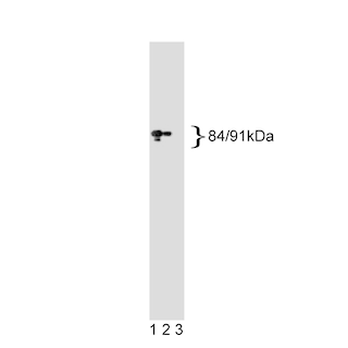
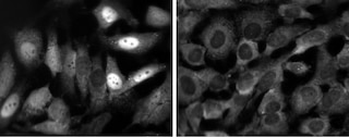
Stat (Signal transducer and activators of transcription) proteins are critical mediators of the biologic activity of cytokines, including interleukins, interferons, erythropoietin, and growth factors. Ligand-receptor interaction leads to activation of constitutively associated JAK family kinases and subsequent recruitment/activation of Stat proteins by tyrosine phosphorylation. Active Stat proteins then move to the nucleus to promote transcription of cytokine-inducible genes. Seven Stat proteins have been cloned, each of which is differentially expressed and/or activated in a cytokine-specific and cell type-specific manner. Stat1 and Stat2 are components of the ISGF3 (Interferon-Stimulated Gene Factor 3) complex, which is the primary transcription activator induced by the binding of the interferon to a specific cell-surface receptor. Stat1 has two alternatively spliced isoforms, 91-kDa Stat1α and 84-kDa Stat1β; Stat1α has 38 additional C-terminal amino acids. In response to the binding of IFNα, IFNγ, EGF, PDGF, or CSF-1 to their respective receptors, the Stat1 subunits become tyrosine-phosphorylated at Y701, and the complex is translocated to the nucleus. This results in the formation of an active complex that includes the DNA-binding p48 subunit. This complex is responsible for modulating the transcription of the interferon-stimulated genes (ISGs).
The 14/P-STAT1monoclonal antibody recognizes the phosphorylated Y701 in Stat1α and Stat1β.
Please refer to Support Documents for Quality Certificates
Global - Refer to manufacturer's instructions for use and related User Manuals and Technical data sheets before using this products as described
Comparisons, where applicable, are made against older BD Technology, manual methods or are general performance claims. Comparisons are not made against non-BD technologies, unless otherwise noted.
For Research Use Only. Not for use in diagnostic or therapeutic procedures.
Report a Site Issue
This form is intended to help us improve our website experience. For other support, please visit our Contact Us page.