







准备和存储


商品通知
- This reagent has been pre-diluted for use at the recommended Volume per Test. We typically use 1 × 10^6 cells in a 100-µl experimental sample (a test).
- Source of all serum proteins is from USDA inspected abattoirs located in the United States.
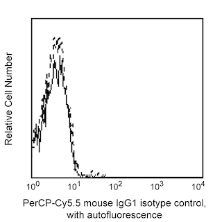
- An isotype control should be used at the same concentration as the antibody of interest.
- Please refer to www.bdbiosciences.com/us/s/resources for technical protocols.
- Cy is a trademark of Amersham Biosciences Limited. This conjugated product is sold under license to the following patents: US Patent Nos. 5,486,616; 5,569,587; 5,569,766; 5,627,027.
- Please observe the following precautions: Absorption of visible light can significantly alter the energy transfer occurring in any tandem fluorochrome conjugate; therefore, we recommend that special precautions be taken (such as wrapping vials, tubes, or racks in aluminum foil) to prevent exposure of conjugated reagents, including cells stained with those reagents, to room illumination.
- Caution: Sodium azide yields highly toxic hydrazoic acid under acidic conditions. Dilute azide compounds in running water before discarding to avoid accumulation of potentially explosive deposits in plumbing.
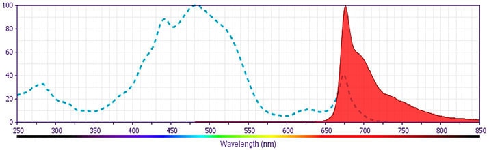
- PerCP-Cy5.5–labelled antibodies can be used with FITC- and R-PE–labelled reagents in single-laser flow cytometers with no significant spectral overlap of PerCP-Cy5.5, FITC, and R-PE fluorescence.
- PerCP-Cy5.5 is optimized for use with a single argon ion laser emitting 488-nm light. Because of the broad absorption spectrum of the tandem fluorochrome, extra care must be taken when using dual-laser cytometers, which may directly excite both PerCP and Cy5.5™. We recommend the use of cross-beam compensation during data acquisition or software compensation during data analysis.
- For fluorochrome spectra and suitable instrument settings, please refer to our Multicolor Flow Cytometry web page at www.bdbiosciences.com/colors.
- This product is subject to proprietary rights of Amersham Biosciences Corp. and Carnegie Mellon University and made and sold under license from Amersham Biosciences Corp. This product is licensed for sale only for research. It is not licensed for any other use. If you require a commercial license to use this product and do not have one return this material, unopened to BD Biosciences, 10975 Torreyana Rd, San Diego, CA 92121 and any money paid for the material will be refunded.
配套商品



The P51-311 monoclonal antibody binds to human BMI-1 (B lymphoma Mo-MLV insertion region 1 homolog). BMI1 is a c-myc cooperating oncogene that encodes an ~45 kDa protein that is a member of the Polycomb Group (PcG) of proteins. PcG proteins are essential for the maintenance, but not initiation, of the transcriptionally repressed state of certain developmental genes. PcG proteins are a structurally diverse group of proteins with conserved functions from fly to human cells. PcG proteins form complexes and regulate the expression of genes involved in cell cycle, DNA repair and differentiation that are crucial for maintaining the self renewal of normal and cancer stem cells. Specifically, BMI-1 is a core component of PRC1 (polycomb repressive complex 1). BMI-1, via the up-regulation of hTERT and independent of c-myc, can immortalize mammary epithelial cells. BMI-1 has also been shown to repress the INK4A locus that controls the tumor suppressors p16 and p19ARF (mouse homologue of p14ARF) in mouse models. BMI-1 plays a role in maintaining the self-renewal capacities of stem cells including hematopoietic, intestinal, retinal and neural stem cells. During antibody development, the purified P51-311 monoclonal antibody was found to detect BMI-1 by Western blot analysis of cellular lysates and by indirect immunofluorescent staining and flow cytometric analysis of fixed and permeabilized cells.
研发参考 (6)
-
Dimri GP, Martinez JL, Jacobs JJ. The Bmi-1 oncogene induces telomerase activity and immortalizes human mammary epithelial cells. Cancer Res. 2002; 62(16):4736-4745. (Biology). 查看参考
-
Itahana K, Zou Y, Itahana Y. Control of the replicative life span of human fibroblasts by p16 and the polycomb protein Bmi-1. Mol Cell Biol. 2003; 23(1):389-401. (Biology). 查看参考
-
Molofsky AV, Pardal R, Iwashita T, Park IK, Clarke MF, Morrison SJ. Bmi-1 dependence distinguishes neural stem cell self-renewal from progenitor proliferation. Nature. 2003; 6961(962):967. (Biology). 查看参考
-
Park IK, Qian D, Kiel M, et al. Bmi-1 is required for maintenance of adult self-renewing haematopoietic stem cells. Nature. 2003; 423(6937):302-305. (Biology). 查看参考
-
Sangiorgi, E., Capecchi, M. R. Bmi1 is expressed in vivo in intestinal stem cells. Nat Genet. 2008; 40(7):915-920. (Biology). 查看参考
-
Tian H, Biehs B, Warming S, et al. A reserve stem cell population in small intestine renders Lgr5-positive cells dispensable. Nature. 2011; 478(7368):255-259. (Biology). 查看参考
Please refer to Support Documents for Quality Certificates
Global - Refer to manufacturer's instructions for use and related User Manuals and Technical data sheets before using this products as described
Comparisons, where applicable, are made against older BD Technology, manual methods or are general performance claims. Comparisons are not made against non-BD technologies, unless otherwise noted.
For Research Use Only. Not for use in diagnostic or therapeutic procedures.
